 Home
Home|
Table of Content Volume 4 Issue 1 -October 2017
Cadaveric study of morphological variations of fissures and lobes of lungs and their clinical significance
Priya P Wattamwar*, Azhar Ahmed Siddiqui**
*Associate Professor, **Professor and HOD, Department of Anatomy, JIIU’s Indian Institute of Medical Science and Research, Warudi, Tq. Badnapur, Dist. Jalna, Maharashtra, INDIA. Email: drpriya2907@gmail.com
Abstract Background: Anatomical variations of lungs which include fissures and lobes of lungs are very necessary for the clinicians for recognizing their variable imaging appearances as well as related abnormalities. Aims and Objectives: So present study was undertaken to note such variations and compare these results with the other studies as well as to identify their clinical significance. Methods: For the study 32 pairs of formalin fixed lungs were taken. These specimens were cleaned, fine dissected and thoroughly observed for any variations of the fissures and lobes of the lungs. Variations were noted and photographed. Results: Among the left lungs, 01 showed presence of incomplete oblique fissure while in another lung there was total absence of oblique fissure, although in one lung there was presence of oblique fissure in the middle part of lung on the sternocostal surface. Accessory fissure was seen in 07 lungs. In right lungs 02 had incomplete oblique fissure, while horizontal fissure was incomplete in 12 lungs. 05 lungs were without the horizontal fissure. Accessory fissure was present in 05 lungs. Conclusion: The present results showed variations on the left side in 35.71% of total lungs in the form of incomplete/absent oblique fissure and presence of accessory fissure. On the right side 75% of the total lungs showed variations in the form of incomplete/absence of fissures and presence of incomplete fissures. These results were compared with previous studies n found to be correlated. Knowledge of these variations is important for both the diagnosis and treatment of various diseases of lungs. Key Words: Oblique, fissure, accessory, lobes.
INTRODUCTION The lungs are the essential organs of respiration. They are situated on both side of heart and other mediastinal contents. Each lung is free in its pleural cavity except for its attachment to trachea and heart at the hilum and pulmonary ligament respectively. The right lung is divided into superior, middle and inferior lobes by an oblique and a horizontal fissure. The upper oblique fissure separates the inferior from the middle and upper lobes and corresponds closely to the left oblique fissure, although it is less vertical and crosses the inferior border of the lung 7.5cm behind its anterior end. The short horizontal fissure separates the superior and middle lobes. It passes from the oblique fissure, near the mid-axillary line horizontally forwards to the anterior border of the lung, level with sternal end of fourth costal cartilage, then passes backwards to the hilum on the mediastinal surface. On a frontal chest radiograph the horizontal fissure is visible in 60% population. The oblique fissure is usually visible in a lateral radiograph and on a high resolution CT as a curvilinear band from the lateral aspect to the hilum. The left lung is divided into a superior and an inferior lobe by an oblique fissure which extends from the costal to the medial surfaces of the lung both above and below the hilum. The left oblique fissure is usually more vertical than the right and is indicated approximately by the medial border of the scapula when the arm is fully abducted above the shoulder. A left horizontal fissure is a normal variant in 10% of patients1. Lung fissures are double fold of visceral pleura that either completely or incompletely invaginate lung parenchyma to form the lung lobes. Accessory fissures of the lung usually occur at the borders of the bronchopulmonary segments. They are common normal variants but are less commonly seen on imaging. Accessory fissures are azygos, inferior accessory, superior accessory, left horizontal and vertical fissure line.
MATERIAL AND METHODS This study was conducted on 64 lungs removed from 32 formaline fixed cadavers from JIIU’s Indian Institute of Medical Science and Research, Warudi, Dist –Jalna and Government Medical College, Aurangabad of Maharashtra state. It included 32 lungs from right side and 28 lungs from left side. The 4 specimens from left side which were damaged during removal or showing pathological lesions or marks of previous surgeries were excluded from the study. The lungs were studied for the following morphological features:
All these features were noted and photographed.
RESULTS In the present study following morphological features were observed. In left lungs: Out of total 28 lungs, 18 lungs were normal showing presence of complete oblique fissure. 02 lungs showed presence of incomplete oblique fissure, while in 01 lung there was total absence of oblique fissure having one lobe only. Presence of incomplete accessory fissure was observed in 07 lungs, and this fissure was most commonly seen in the superior lobe of the left lung (in 05 lungs). In right lungs: Total 32 right lungs were studied. Presence of normal and complete oblique as well as horizontal fissure was seen in 08 lungs. Incomplete oblique fissure was present in 02 lungs while incompleteness of horizontal fissure was seen in 12 lungs. The horizontal fissure was absent in 05 lungs. 05 lungs showed presence of incomplete accessory fissure which was present in the inferior lobe.
Table 1: Showing findings in left lung (n=28)
Table 2: Showing findings in right lung (n=32)
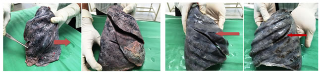
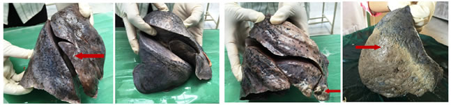
The nature of the fissure is of great importance in planning operative strategy for thoracoscopic pulmonary resection where an incomplete fissure may contribute to post operative leak. As no method of clear classification of pulmonary fissures was present, Craig and Walker2 proposed an anatomical classification of the pulmonary fissures based on both the degree of completeness of the fissures and the location of the pulmonary artery at the base of the oblique fissure. Four stages have been described: Grade I: Complete fissure with entirely separate lobes; Grade II: Complete visceral cleft but parenchyma fusion at the base of the fissure. Grade III: Visceral cleft evident for a part of the fissure; Grade IV: Complete fusion of lobes with no evident fissure line. DISCUSSION Development of the lungs begins on the 22 day with formation of a ventral out pouching of the endodermal foregut called the respiratory diverticulum. This bud grows ventrocaudally through the mesenchyme surrounding the foregut and on days 26 to 28 it undergoes a first bifurcation splitting into primary right and left primary bronchial buds. Many lung anomalies result from a failure of the respiratory diverticulum or its branches to branch or differentiate correctly. Errors in the pattern of pulmonary branching during the embryonic and early fetal periods result in defects ranging from an abnormal number of lobes or bronchial segments to the complete absence of a lung. Anomalies of lobation of the lungs may be produced by fusion of adjacent lobes to obliterate a fissure, by occurrence of abnormal fissures, or by the aplasia or agenesis of a part of a lung3. During development fissures separate individual broncho-pulmonary segments as the lungs grow. The spaces remain along the interlobar planes to give rise to major and minor fissures in a fully developed lung. Absence or incompleteness of a fissure could be due to obliteration of these fissures either completely or partially. With defective pulmonary development there can be variations in the lobes and fissures of lung4. Lung fissures are double fold of visceral pleura that either completely or incompletely invaginates lung parenchyma to form the lung lobes. Anatomically an accessory fissure is a cleft of varying depth lined by visceral pleura. These fissures vary in depth from one individual to another, but never completely separate the lobes at the hilum, where there is some fusion between adjacent lobes; in a surgical resection of a lobe the fissures must therefore be extended by dissection to the hilum5. The human lung was first characterized as asymmetrical by Ae by because of the presence of a right eparterial bronchus. Presence of supernumerary fissures is the most common variation of lungs. These fissures do not always separate segments but may enter subsegmental or interbronchial planes. Another common form of lung variation includes the absence of fissures. In a study of 277 lungs, the horizontal fissure was absent in 21% and incomplete in 67%. Incomplete oblique fissures occur in about 30% of both right and left lungs6. Lymphatics of lung drain centripetally from pleura towards the hilum. Altered course of oblique fissure would mean altered course of visceral pleura, thereby changing the arrangement of lymphatic drainage7. Figure 1 Figure 2 Figure 3 Figure 4 Figure 5 Figure 6 Figure 7 Figure 8 Figure 1: Showing incomplete accessory fissure of left lung; Figure 2: Showing accessory fissure of right lung; Figure 3: Showing absence of horizontal fissure with fissure of right lung; Figure 4: Showing incomplete oblique fissure of incomplete oblique left lung; Figure 5: Showing incomplete horizontal fissure; Figure 6: Showing hidden middle lobe of right lung of right lung; Figure 7: Showing accessory lobe of right lung; Figure 8: Showing absence of oblique fissure in left Lung Table 3: Showing Comparative prevalence of anatomical cadaveric studies
A Fetal study was also done on 61 formalin fixed human fetuses ranging from 12 – 40 weeks of gestational age. In this study presence of accessory fissures was more on right side than the left side. Prevalence of superior accessory fissure was 14.5% in right side with absence in left lung. Inferior accessory fissure was not found. While Left minor fissure prevalence was 12.9%18. Autopsy studies have also been done on lungs to access the fissures of the lungs. Nadire19 et al studied morphologic and morphometric features of major fissures and major accessory fissures in 420 Anatolian autopsy lungs. In this study they observed presence of major accessory fissure most commonly in the inferior lobe of the left lung. In Bates study20 which was performed on fetuses with anomalous fissures it is reported that other malformations were approximately three times more common than in fetuses with a normal fissure pattern. It has been reported in the literature that the true lobar pattern abnormalities occur in some conditions, notably sinus inversus viscerum, polysplenia and asplenia, where it is believed to be an underlying defect in the normal process of determination of the right left asymmetry. Polysplenia syndrome is characterized by bilateral bilobed lungs and asplenia by bilateral trilobed lungs; these syndromes are sometimes referred to as bilateral left or right sidedness respectively. Various radiographic studies have also been done to evaluate main as well as accessory fissures of the lungs. Most prevalent abnormality found was that of absent or incomplete horizontal fissure of right lung. Inferior accessory fissure was the most common accessory fissure found in these studies. Yildiz21 et al found the inferior accessory fissure as the most common (12%) variant in its study, with left minor fissure (8%) being the second most common followed by right superior accessory fissure (5%). While Kilic22 et al reported right inferior accessory fissure as the most common accessory fissure. In the present study incomplete or absent horizontal fissure of right lung was the most common found abnormality and the inferior accessory fissure was being found in 2 of the left lungs and 5 of the right lungs. Superior accessory fissure was mostly seen in the left lungs only (05 lungs). Table 4: Showing Comparative prevalence of CT studies
According to Travor27 accessory fissures of the lung are commonly observed in lung specimens but are often unappreciated or misinterpreted on radiographs and CT scans. They usually occur at the boundaries between bronchopulmonary segments. Most common are the inferior accessory fissure which demarcates the medial basal segment, the superior accessory fissure which demarcates the superior segment and left minor fissure which demarcates the lingual. Radiographically it appears as a thin white line resembling the major or minor fissure except for location. This line can be mistaken for an interlobar fissure for a scar, for the wall of a bulla or for a pleural line made visible by pneumothorax. Besides a thin line the other usual radiographic manifestation of an accessory fissure occurs when the fissure acts as a barrier to the spread of infection, creating a sharply marginated pneumonia that can be mistaken for at electasis or consolidation of a whole lobe or even for a mediastinal mass. Also these incomplete fissures may alter the usual patterns of collapse seen in patients with endobronchial lesions. These can give rise to the unexpected appearances of pleural effusions. An incomplete major fissure causes an odd appearance of fluid tracking within the fissure. Where both minor and major fissures are incomplete the classic middle lobe stair step of pleural effusion is seen. Incomplete fissures may also alter the spread of the disease within the lung. Because of the fissures, pneumonia in a particular lobe is often limited to that lobe alone. In patients with incomplete fissures pneumonia may spread to adjacent lobes through the incomplete fissures. Odd lobar involvement with carcinoma of the lung may be explained on a similar basis. Additional fissures are thus most common variant in pulmonary development. They are more frequent in fetal and neonatal lung specimens than in adults28. The identification of the completeness of the fissures is important prior to the lobectomy, because individuals with incomplete fissures are more prone to develop post-operative air leak, and may require further procedures such as stapling and pericardial sleeves. These air leaks can be avoided by the surgeon by properly clamping the fused pulmonary lobe segments29. Also it is important to identify the incomplete fissures, as it is believed that they may allow collateral ventilation between the lobes which may compromise lobar exclusion30. Knowledge of anatomy and variations of the fissures and lobes of lungs is important for the radiological imaging as well as other associated abnormalities. Also this will reduce the peri-operative complications in surgeries involving lungs. CONCLUSION This study was done to assess the normal fissures and lobes of the lungs as well as to know the variations in these morphological features of the lungs. It is seen that incomplete or absent horizontal fissure was the most common abnormality observed in this study, followed by presence of accessory fissures; inferior being common in right lungs while in left lungs superior accessory fissure was more common. Cadaveric studies are also important to know the prevalence of the abnormalities associated with the fissures and lobes of the lungs. This will help the radiologists as well as surgeons planning lobectomies and segmentectomy.
REFERENCES
|
|
 This work is licensed under a Creative Commons Attribution-NonCommercial 4.0 International License.