 Home
Home|
Table of Content Volume 9 Issue 2 - February 2019
Bilateral variations in the terminal branches of facial nerve - A case report
Alka Bhingardeo1*, Mehera Bhoir2, Tejas Shelar3
1Registrar, 2Head, 3MBBS Student, Department of Anatomy, HBTMC and Dr. R. N. Cooper Hospital, Mumbai, Maharashtra, INDIA. Email: dr.alkabhingardeo@gmail.com
Abstract Facial nerve is the seventh cranial nerve. It supplies the muscles of facial expression. It gives five terminal branches after emerging from the parotid gland. The functional conservation of facial nerve through proper detection in surgeries around parotid region and procedures like facial rhytidectomy is necessary. Facial nerve paralysis is a stressful complication of parotid surgery. To avoid all the postoperative morbidity associated with the injury to the facial nerve, detailed knowledge of the branching pattern of the facial nerve and its probable variations is must. Current article is about several communications between the terminal branches of the facial nerve leading to the different branching pattern on the right and the left side found during routine cadaveric dissection in an adult male cadaver. Key Word: facial nerve, parotid surgery, facial nerve paralysis.
INTRODUCTION Facial nerve is the seventh cranial nerve. In its extracranial course, after emerging from the stylomastoid foramen, normally the facial nerve enters the parotid gland and passes between superficial and deep part of the gland winding around the isthmus. It divides into two trunks – temporofacial and cervicofacial inside the gland. Temporofacial trunk further divides into the temporal and zygomatic while cervicofacial trunk divides into the buccal branch, marginal mandibular and cervical branches1,2,3,4 Normally there is no anatomic communications between these terminal branches of the facial nerve that supply the variousmuscles of face. In this cadaver, we found various communications between terminal branches of the facial nerve leading to the different branching patterns on the right and the left sides. Preserving emerging branches of facial nerve is the matter of major concern to the surgeons during parotid surgery to avoid postoperative morbidity. Therefore, appreciation of relative anatomy of the facial nerve and knowledge of its associated variations is important.5,6
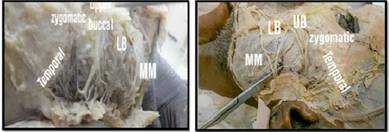
CASE REPORT During routine cadaveric dissection in an adult male cadaver, we found variations in terminal branches of facial nerve. Facial nerve in this case divided into two trunks temporofacial and cervicofacial. But further branching pattern was different than normal on right and left sides. On right side, we found there was ramification of the temporal branch into six divisions. Buccal branches were two upper buccal, above the parotid duct and lower buccal, below the parotid duct. The Zygomatic branch was single and there was communication between zygomatic and upper buccal branch. Upper buccal branch was coming from two roots. Lower buccal was a single branch and there was communication of lower buccal not only with upper buccal but also with marginal mandibular branch. Marginal mandibular branch was single and found below the lower buccal. It joined lower buccal branch and this union passed upward and formed a loop with the upper buccal branch. Such type of migration of marginal mandibular upwards forming a loop with the other branches is called as plexiform arrangement as per Devis6 classification. Cervical branch was single and crossed the angle of mandible and supplied platysma.On the left side, temporal branch was represented by five divisions like that of right side but the variations in the other branches were different. The zygomatic branch was originating from the common stem with the upper buccal branch and further it divided into three divisions. Upper buccal was arising from a common stem with the zygomatic. It passed above the parotid duct and formed a loop with the lower buccal after travelling some distance. Lower buccal branch was found to arise from a common stem with the marginal mandibular. It was a single branch and formed a loop with upper buccal branch. Marginal mandibular was a single branch in common stem with the lower buccal and passed below the lower buccal branch. Cervical branch on left side found to have same normal course as that like on the right side without any variation. So to sum up there were variations of communication within branches of temporofacial trunk as well as cervicofacial trunk. The variations were different on the right and left side. Variation in the branching pattern of facial nerve on the right side - Variations in branches of temporofacial trunk on right side -
Variations or communications between branches of cervicofacial trunk on right side
Variation in the branching pattern of facial nerve on the left side – Variations in branches of temporofacial trunk on left side –
Variations or communications between the branches of cervicofacial trunk on the left side -
Figure 1: Branches of facial nerve on right side, Figure 2: Branches of facial nerve on left side
Figure 3: Loop formed by upper buccal, lower buccal and marginal mandibular branch of facial nerve on right side, Figure 4: Loop formed by upper and lower buccal branch of facial nerve on left side
DISCUSSION Variations in the branching pattern of facial nerve are not uncommon. We found that temporal branch was represented by five rami on both the sides. Gosain A K7, mentioned in his article that as per recent studies temporal branch is not a single branch but represented by more than one branches. We observed that on the right side there was a single zygomatic branch which was arising from the two rami and ran between lowermost ramus of temporal and upper buccal. This zygomatic nerve also showed communication with the upper buccal. The communication was H shaped. While on the left side zygomatic branch originated from a common stem with the upper buccal branch. In our study, we found two buccal branches upper buccal above the parotid duct while lower buccal below the parotid duct on both right and left sides. Liu AT8 in his study on facial nerve also found two buccal branches in 87.5% of cases. We found communication of upper buccal with the lower buccal. In addition to this on right side upper buccal was also communicating with the zygomatic branch while lower buccal was communicating with marginal mandibular branch while on left side there was common stem of upper buccal with zygomatic while of lower buccal with marginal mandibular. In addition to this on left side there was a loop of communication between the upper buccal and lower buccal around the parotid duct. Gosain A K7 said in his article that interconnections between the zygomatic and buccal branches were found in more than 70% of cases. In the study of such anastomotic patterns of facial nerve, Bandell H9 mentioned that earlier studies found communication between temporofacial and cervicofacial trunks in 60to 70 % of cases where as he found such communications in only 44% of cases. Lower rate of occurrences of such connections between the two trunks result in increased risk of facial palsy after transection of terminal branch. Temporal and zygomatic branches are more prone to injury in procedures like facial rhytidectomy, coronal or endoscopic bro lifting and temporal craniotomy.1 In our case, we found marginal mandibular nerve was represented by a single branch on both the sides. On the right side it was communicating with the lower buccal branch. This communication formed a loop with the upper buccal branch. Variations in the marginal mandibular branch is not uncommon. Karapinar U10 et al in his study on the course of nerve found that marginalmandibular nerve was represented by single branch in 36.4% of cases while he found two marginal mandibular nerve divisions in 63.6 % of cases. The author also found the communication of mandibular branch with the buccal branch in 4.6% of cases but had not described the type of communication. Woltmann M11 in his study also found communication of marginal mandibular branch not only with the buccal but also with the cervical branch.In surgeries of submandibular region, there are chances of injury to the marginal mandibular branch. Any injury to this nerve during the operation results in significant post–operative morbidity12. Saylam C13 mentioned in his article that marginal mandibular nerve is one of the most vulnerable branches to the surgical injury because of its location and surgeons operating specially for rhytidectomies should have a true knowledge of this branch.
CONCLUSION The communications within branches of facial nerve are important during clinical examination and surgical procedures of facial nerve. In the surgical repair of facial nerve paralysis, a tension free end to end coaptation of the trunk or its branches is important.14 Knowledge of such variations in the branching pattern of facial nerve is significant in facial nerve reconstructive surgery, neck dissection and in various nerve transfer procedures. Information regarding such variations may also help in understanding the pathophysiology of disorders related to the nerve15.
REFERENCES
|
|

 This work is licensed under a Creative Commons Attribution-NonCommercial 4.0 International License.