 Home
Home|
Table of Content Volume 11 Issue 1 - July 2019
Morphometric study of human adult cadaveric spleen
Manisha S More1, Manoj D Togale2*, Shilpa Bhimalli3
1Associate Professor,3Professor and Head, Department of Anatomy, Jawaharlal Nehru Medical College, KLE Deemed to be University, Belagavi, Karnataka, INDIA. 2Associate Professor, Department of Surgery Jawaharlal Nehru Medical College and KLES Dr. Prabhakar Kore Hospital, KLE Deemed to be University Belagavi, Karnataka, INDIA. Email: zapmanojtogale@yahoo.com , manishamtogale@yahoo.co.in Abstract Background: Spleen is a hemolymph organ in the human body. It is the largest and clinically important lymphoid organ. It can show a wide range of variation, the knowledge of which is important for physicians, surgeons and radiologists Aim of the Study: The present study was done to perform a morphometric analysis of human cadaveric spleens and compare the results with previous studies. Materials and Methods: The present study was done on 41 human adult cadaveric spleens obtained from Department of Anatomy, J.N. Medical College, Belagavi. The morphological features of the spleen like its length, breadth, width and weight were measured. The shape, poles, borders, surfaces and the impressions on the spleen were observed Results: The lengths of the spleens varied between 6.8 cm to 14cm, with an average of 9.97 cm. Their breadth was between 5.20and 10.4cm. The average breadth was 7.21 cm. Their widths varied between 2.1 and 6.4 cm, with an average of 3.65 cm. The weights of the spleens showed great variations, ranging between 51.70 and 369.90 gm, with an average of 132.79 gm. Various shapes of the spleens were observed in the present study. Most of the spleens were wedge shaped [48.78%], followed by tetrahedral [14.63%] and triangular [17.1%]. Additional oval [7.32%], semilunar [7.32%], heart shaped [2.44%], and irregular shapes [2.44%], of the spleens were observed. In all the spleens, two poles, two borders and two surfaces were observed. The diaphragmatic surface of the spleen showed a uniform morphology while its visceral surface showed gastric, renal, colic and pancreatic impressions. The splenic notches were present on the superior as well as on the inferior borders. In most of the cases [85.37%], the notches were found on the superior border. The number of notches varied from zero to six, but in most of the cases [31.71%], there were 1 notch. Conclusion: These findings will be helpful for operating Surgeons and interventional Radiologists and for Anatomists too. Key Word: Spleen, shape of spleen, spleen notches.
INTRODUCTION Spleen is the important hemolymph organ in humans. It is largest lymphoid organ in the body. It is situated in the upper left hypochondriac region and partly in the epigastrium. It is covered from all the sides by peritoneum and is closely related to the fundus of the stomach, left kidney, left colic flexure and the diaphragm. It has two poles, i.e. anterior and posterior, two surfaces i.e. diaphragmatic and visceral, and three margins i.e. superior, inferior, and intermediate. It’s shape varies from a slightly curved wedge to a domed tetrahedron1. The size and weight of the spleen varies with age1. On an average it is 12 cm long,7 cm broad and 4 cm thick in the adult. Weight of the spleen ranges from 80 to 300 g average being 150 g.1,2,Splenomegaly is important diagnostic sign in malaria, kalaazar, inflammatory and degenerative disorders3 . In splenomegaly, the anterior border, anterior part of diaphragmatic surface and notched superior border may become clearly palpable below the left costal margin4 Spleen begins to develop during 5th week of intrauterine life from a mass of mesenchymal cells originating in the dorsal mesogastrium as a localised thickening of coelomic epithelium.5 The spleen is nodular in fetus, but the lobules normally disappear before birth. The notches in the superior border of the adult spleen are remnants of the grooves that seperated fetal lobules.5 Spleen assumes clinical importance due to hematological and immunological role. Surgeons also like to conserve splenic tissue during splenectomy due to the same reasons. The present study was undertaken to describe the morphometric variations in spleen and compare it with the available literature which would prove useful to both clinicians and academicians. MATERIALS AND METHODS This study was done on 41 formalin fixed human adult cadaveric spleens of both sexes obtained during routine dissection classes of undergraduate medical students in Anatomy department of J. N. Medical College, Belagaviand USM-KLE University, Belagavi. The morphological features of the spleen which includes its length, breadth, width (mm) and weight (gms) were measured by using digital Vernier caliper and electronic weighing machine respectively. The shape, poles, borders, surfaces and the impressions on the spleen were observed. The notches on the borders of the spleen were observed carefully. Pictures were taken wherever necessary. As in the study which was done by Michels et al6, Length (mm)- the greatest distance between the two poles of the spleen, Breadth (mm)- the greatest distance between two points at the same level on the superior and inferior borders and Width (mm)- the greatest width of the spleen. RESULTS Measurements of 41 spleens are shown in table-1 Table 1: showing measurements of 41 spleen
In the present study, out of 41 spleens, 20(48.78%) were wedge shaped,7( 17.1%) were triangular shaped, 6 (14.63%) were tetrahedralshaped,3(7.32%) were oval shaped ,3 (7.32%)semilunar shaped,1(2.44%) heart shaped and 1 (2.44%) were irregular in shape. (table 2)
Table 2: variations in shapes of spleens
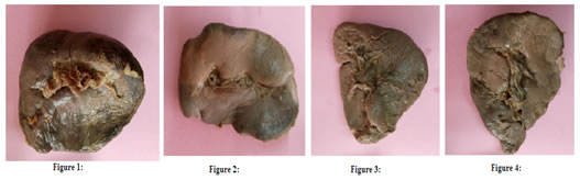
Figure 1: Wedge shape of spleen; Figure 2: Tetrahedral shape of spleen; Figure 3: Triangular shape of spleen; Figure 4: Triangular with tongue shaped projection shape of spleen
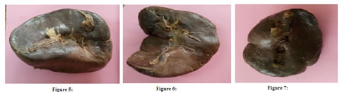
Figure 5: Semilunar shape of spleen; Figure 6: Heart shaped spleen; Figure 7: Oval shape of spleen.
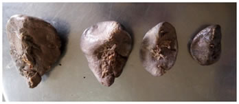
Figure 8: showing changes in size of spleen Weight of 41 spleens ranged between 45.5 to 369.90gm, with an average of 132.79 gm. The maximum number of specimens i.e. 22 (53.66%) have weights in the range of 80 to 150 gm. (table 3)
Table 3: variations in weight of spleens
In the present study, the lengths of the spleens varied between 6.8 cm and 14 cm, with an average length of 9.97 cm.Most of the spleens were in the range of 10 cm to 12 cm in length (43.9%) (Table 4)
Table 4: variations in length of spleens
The breadth was spleen varied between 5.2 cm and 10.4 cm, with an average breadth of 7.21 cm. Most of the spleens were in the range of 5.6 cm to 7.5 cm in breadth (56.12%) (Table 5)
Table 5: variations in Breadth of spleens
It was found that the widths of the spleens varied from 2.1 cm to 6.4cm with an average width of 3.65 cm. In most of the cases spleens width was in the range of 2 cm to 4 cm (58.54%). (table 6) Table 6: variations in Width of spleens
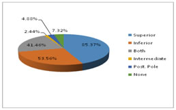
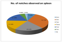
The splenic notches were present on the superior as well as on the inferior borders. In most of the cases [85.37%], the notches were found on the superior border. The number of notches varied from zero to six, but in most of the cases (31.7%),there were 1 notch.(table 7 and 8)
Table 7: borders of spleen showing presence of notches
Figure 1: borders of spleen showing presence of notches
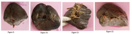
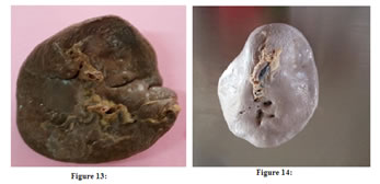
Figure 9: showing notch on both inferior order; Figure 10: showing notches both superior and inferior order; Figure 11: showing multiple notches on superior border; Figure 12: showing notch on posterior pole; Figure 13: showing notch on inferior border; Fig.14- showing no notch Table 8: No. of notches present on spleen
Graph 2: Number of notches observed on spleen Table 9: showing mean values of measurements of spleen
Table 11: Variation in shapes of spleen
Table 12: variations in notches of spleen
DISCUSSION The spleen is an important haemolymph organ. As reported by Michels6 and as mentioned in Gray’s anatomy1, in the present study also, so many variations were found in the morphology of the spleen. The values for the length, breadth, width and weight of the spleen in the present study were slightly lower than previous studies done in western countries compared to Indian studies.1,6,7,8,9,10 This may be due to the differences in the genetic factors, body constitution, geographical conditions, feeding habits and the better socioeconomic status, in the western countries where these studies were done. Presence of Notches on spleen- The spleen develops from the mesoderm. During its development, different lobules are formed, which fuse with each other later on. The indication of the lobulation in adult spleen is its notchedupper border.11 Sometimes, this lobulated appearance may persist in the spleen which indicates improper fusion of spleen and causes many notches on the spleen, which can be seen most commonly on the superior as well as on the inferior borders. In the present study, the splenic notches were found on the superior as well as on the inferior borders. The number of notches varied from zero to six, but commonly, there were only one or two notches. These findings of the study in accordance with previous studies.2,6 Presence of notches on the superior margin is useful for the physician to palpate the spleen during enlargement of spleen9,12. Presence of notches in the inferior border may be important for surgeons attempting splenic surgeries and radiologists interpreting CT scans.
CONCLUSION The knowledge on the anatomical variations of the spleen is of fundamental importance to the clinicians during the routine clinical examinations of the abdomen, to the surgeons while they perform surgical procedures which are related to the spleen, to the radiologists for their diagnostic procedures and of course The detailed knowledge on spleen is important to avoid and prevent any complications and to obtain a good operative, as well as diagnostic intervention. This knowledge is also very important for anatomists during their routine dissection procedures. REFERENCES
|
|
|||||||||||||||||||||||||||||||||||||||||||||||||||||||||||||||||||||||||||||||||||||||||||||||||||||||||||||||||||||||||||||||||||||||||||||||||||||||||||||||||||||||||||||||||||||||||||||||||||||||||||||||||||||||||||||||||||||||||||||||||||||||||||||||||||||||||||||||||||||||||||||||||||||||||||||||||||||||||||||||||||||||||||||||||||||||||||||||||||||||||||||||||||||||||||||||||||||||||||||||||||||||||||||||||||||||||||||||||||||||||||||||||||||||||||||||||||||||||||||||||||||||||||||||||||||||||||||||||||||||||||||||||||||||||||||||||||||||||||||||||||||||||||||||||||||||||||||||||||||||||||||||||||||||||||||||||||||||||||||||||||||||||||||||||||||||||||||||||||||||||||||||||||||||||||||||||||||||||||||||||||||||||||||||||||||||||||||||||||||||||||||||||||||||||||||||||||||||||||||||||||||||||
 This work is licensed under a Creative Commons Attribution-NonCommercial 4.0 International License.