 Home
Home|
Table of Content Volume 11 Issue 1 - July 2019
The study of nutrient foramina of tibia in Marathwada region of Maharashtra
R D Walwante1, S S Dhapate2*, K Thomas Manoj3
1,3Assistant Professor, 2Professor and HOD, Department of Anatomy, SRTRGMC, Ambajogai, Dist. Beed, INDIA. Email: drdhapatess@gmail.com
Abstract Background: Nutrient foramen is a natural opening into the shaft of a bone, allowing for passage of blood vessels into the medullary cavity. This study aims to determine the number, location and direction of nutrient foramina of tibia. Material and methods: 185 dry tibia from government medical colleges from Marathwada region were examined for nutrient foramina location number direction and its relation to the other bony land marks. Results: Single nutrient foramina directed downwards was the rule. The location of nutrient foramina closely resembled with the data of previous similar studies done in other part of world. Conclusion: The present study generated database for nutrient foramen of tibia from Marathwada area. This can be utilized by surgeon to preserve nutrient artery during operation for fracture repair, tumor and bone grafts involving tibia. Key words: Tibia, Nutrient artery, Nutrient foramen, Fracture repairs.
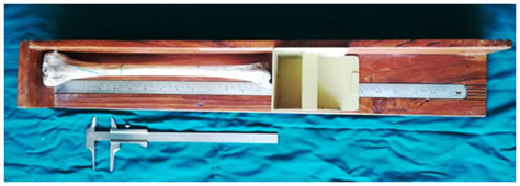
INTRODUCTION Nutrient foramen is an opening in the bone shaft which gives passage to the blood vessels of the medullary cavity of a bone for its nourishment and growth1. The nutrient artery is the principal source of blood supply to a long bone and is particularly important during its active growth period in the embryo and fetus, as well as during the early phase of ossification2. The nutrient artery is supplemented by epiphyseal, metaphysical and periosteal vessels in providing blood supply to the long bones. The diaphyseal nutrient arteries obliquely penetrate in the diaphysis of the long bones, their entrance point and angulations being relatively constant, dividing in ascending and descending branches, once they reach the medullary cavity3. It has been suggested that the direction of the nutrient foramina is determined by the growing end of the bone. The growing end is supposed to grow at least twice as fast as the other end. As a characteristic, the nutrient vessels move away from the growing end in the bone4.The nutrient artery to the tibia is a branch of posterior tibial artery or peroneal artery. It supplies the inner two-thirds of the cortex and is the chief blood supply of cortical bone5. The nutrient artery enters the shaft of the tibia generally in its upper one third; this arrangement deprives the nutrition to the lower one third of shaft, thus making the lower one third of tibia more liable to undergo nonunion in the event of fracture6.A considerable interest in studying nutrient foramina resulted not only from morphological, but also from clinical aspects. The knowledge of location of nutrient foramen in long bones is of importance to the surgeons as minimal interference with the vascularity of the bone during surgical procedures or during fracture repairs will improve the surgical outcome substantially7-10. Some pathological bone conditions such as developmental abnormalities, fracture healing or acute hematogenic osteomyelities are closely related to the vascular system of the bone11. Detailed data on the blood supply to the long bones and the association with the areas of bone supplied has been continued to be a major factor in the development of new transplantation and resection techniques in orthopedics2, 12. Present study was undertaken to generate the data on exact location of nutrient foramen in tibia from marathwada region and to compare the findings with the similar studies done in other parts of the world. MATERIAL AND METHODS The present study was conducted on dry specimen of tibia from the department of Anatomy of medical colleges from marathwada region. Side determination of the dry tibia was done using existing universally accepted criteria. The Sliding Vernier caliper and osteometric board was used for all the distance measurements.
Figure 1: Shows sliding vernier caliper and osteometric board. Following observations were made on each tibia:
All the observations were tabulated and analyzed by using SPSS software version 20.
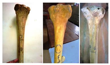
RESULTS The present study was done on 185 dry tibias (87 Right and 98 Left) of both the sexes from the department of Anatomy of medical colleges from Marathwada region. In all the samples nutrient foramen was found on the posterior surface of the shaft out of which single nutrient foramina (NF) was found in 177 samples, double NF was found in 7 samples and one sample is having three NF. Shown in figure 2. Figure 2: Shows number and direction of nutrient foramina on tibia We noticed that in 90 tibias (48.65%) the nutrient foramen was located on the shaft in its upper one third, in 32 tibias (17.30%) it was positioned in middle one third of the shaft and in 63 tibias (34.05%) it was positioned at junction of upper and middle 1/3 of the shaft (Table 1). In all 185 tibias (100%) the direction of nutrient foramen was observed to be downward towards its lower end. The location of nutrient foramen when compared with Soleal line, we determined that in 168(90.81%) tibia it was located lateral to the Soleal line and in 17(9.18%) tibia it was located on the Soleal line (Table 2). Table 1: Location of nutrient foramina in right and left tibia
Table 2 : Direction and relation of nutrient foramina (NF) with the soleal line (SL) on right and left tibia.
In the remaining tibias, the exact distance of the nutrient foramen was measured from two landmarks: i) the junction of upper and middle one third of the shaft; ii) the Soleal line. We discovered that (Table 3) the nutrient foramen was located on posterior surface of shaft: i) In right tibias at an average distance of 9.2 mm ± 8.6 above the junction of upper and middle 1/3rd of the shaft and 10.7mm ± 5.1 is mean distance of NF From Soleal Line. In left tibias at an average distance of 7.3mm ± 7.4 above the junction of upper and middle 1/3rd of the shaft and 10.8 mm ± 5.4 is mean distance of NF from Soleal Line.
Table 3: Distance of nutrient foramina from junction of upper 1/3 and middle 1/3 of shaft and from soleal line on right and left tibia.
Table 4: Comparison with previous studies for location of nutrient foramina of tibia.
Table 5: Comparison with previous studies for relation of nutrient foramina of tibia with soleal line.
DISCUSSION The nutrient artery to tibia is derived from posterior tibial artery near its origin. It is one of the largest of the nutrients arteries. A single nutrient foramen on the shaft of the tibia is a common observation of the past studies. Few researchers have also reported the double diaphysial nutrient foramen on the tibia as a rare occurrence12.In the present study, a single nutrient foramen on shaft was a rule in all 177 tibias with rare exceptions of double NF in 7 tibias and triple nutrient foramina in one tibia. All of these nutrient foramina were directed downwards towards the lower end of the tibia. The nutrient foramen was located in upper one third of tibial shaft in 48.65% and 34.05% at junction of upper and middle 1/3 of shaft of tibia. Thus our study shows that the data from marathwada region on location of nutrient foramen of tibia is not in concurrence with many previous studies done in the different parts of the world, it shows there is increased percentage of location of nutrient foramina at the junction of upper and middle 1/3 of shaft of tibia. Regarding the middle 1/3 location this study is in concurrence with Mysorkar (1967) study. (Table 4)3, 4.12-16. The nutrient foramen was situated lateral to Soleal line >90% of the present study samples. None of the nutrient foramen was lying medial to Soleal line. This finding about relation of nutrient foramen with Soleal line reaffirms the observations made by previous researchers (Table 5).3, 13, 16-18 The knowledge of exact location of nutrient foramen of tibia is highly useful for the surgeons who perform fracture repair or are involved in surgical interventions of tibia. So we measured the distance of nutrient foramen from junction of upper and middle 1/3rd of the shaft and the Soleal line. For this the tibias with nutrient foramen in upper one third of shaft were only included. The findings are shown in Table 3. This data will enable the surgeons to efficiently avoid the damage to the nutrient artery of tibia during surgical procedures involving this region which can have significant impact on overall recovery of patient. So knowledge of nutrient foramen is important for orthopaedic surgeons preoperatively to preserve the circulation in open reduction of fractures, joint replacement therapies and in bone graft surgeries.
CONCLUSION The present study reconfirms that the nutrient artery to tibia enters into its shaft on its posterior aspect lateral to Soleal line in > 90% of the cases but regarding location of nutrient foramina, it was located in upper one third of tibial shaft in 48.65% and 34.05% at junction of upper and middle 1/3 of shaft of tibia. Clinical procedures involving this area shall be done with extreme care to preserve the chief blood supply of tibia. The data from marathwada region of Maharashtra about number, direction and location of nutrient foramen of tibia little bit variations with the data from other parts of the world.
REFERENCES
|
|
||||||||||||||||||||||||||||||||||||||||||||||||||||||||||||||||||||||||||||||||||||||||||||||||||||||||||||||||||||||||||||||||||||||||||||||||||||||||||||||||||||||||||||
 This work is licensed under a Creative Commons Attribution-NonCommercial 4.0 International License.