 Home
Home
|
Table of Content Volume 11 Issue 2 - August 2019
Descent of testes and role of gubernaculum
Vaibhav V Phad1*, R A Joshi2
1Assistant Professor, Department of Anatomy, Shri Vasanatrao Naik Government Medical College, Yavatmal 445001. Maharashtra, INDIA. 2Professor and Head, Department of Anatomy, Government Medical College, Miraj, Sangli 416410. Maharashtra, INDIA. Email: drvaibhavphad@gmail.com
Abstract Background: Gubernaculum testis was described way back in 1762 since then it had been considered to have a role in descent of testis. We have studied its morphology to through light on its role in descent of testis. Materials and methods: aborted male human foetuses between 12th to 40th weeks of gestational age with no obvious congenital abnormality were taken for study. The position of testis, length of gubernaculum and its distal attachments of were noted. Observations and results: the length of gubernaculum was increased just before descent and decreased with descent. Also the thickness of gubernaculum was increased during descent. The length remained more or less constant from 12th week to 40th week and a slight decrease in length was observed. Conclusion: the gubernaculum is important structure in descent of testis and along with several other factors helps in descent of testis. Keywords: Gubernaculum, descent of testis, length of gubernaculum, thickness of gubernaculum, ligaments of lockwood.
INTRODUCTION The gubernaculum is a Latin word for helm or rudder. In 1762 John Hunter was the first to publish a detailed description of the structure which "connects the testis with the scrotum and directs its course in its descent", which he named the gubernaculum1. In human foetuses the Testes develop in relation with abdomen and descent with time to their real position in scrotal sac, traversing the abdominal wall between the 15th and 28th weeks of gestation (17th to 30th menstrual weeks). Complications that adversely affect this displacement may lead to cryptorchidism and other testicular abnormalities. However, the mechanisms that regulate testicular migration are not yet well established. Number conflicting theories have been proposed to explain the mechanism of testicular descent. All these theories conclude that the testis was either pulled or pushed from the abdomen into the scrotum, by a combination of growth or involution processes. The most accepted theories for explaining testicular descent in humans are related to increased intra-abdominal pressure(2,3,4,5,6), the development of the epididymis, vas spermatic, vas deferens and inguinal canal7, development of the gubernaculum(8,9), stimulus from the genitofemoral nerve9 and various stimuli from hormones and biologically active peptides with systemic and/or paracrine effects10. In this study the Descent of Testes and Gubernaculum is studied in detail.
MATERIAL AND METHODS Forty aborted male human foetuses between 12- 40 weeks of gestational age (Intra-Uterine Life, IUL) with no obvious congenital anomalies were obtained from the Department of Obstetrics and Gynaecology, GMC, Miraj, and PVPGH, Sangli, with the prior permission of Head of Department and consent of parents. The study was approved by the Ethical Committee. Gestational age, sex, weight and crown- rump length were noted in detail. After taking a median vertical incision on the anterior abdominal wall, the abdominal cavity was opened. Intestines were pushed aside to expose the posterior abdominal wall. The testes were seen along the posterior abdominal wall covered by peritoneum in lower gestational age foetuses. The position of Testes and gubernaculum was noted. The lengths of gubernaculum were noted with calliper. If the testes were in scrotal sac in higher gestational age foetuses the length of scrotal ligament was noted.
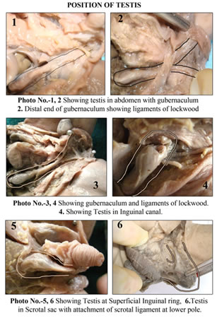
OBSERVATIONS AND RESULTS Positions of testes were noted according to the Groups of Gestational Age in weeks, Weight in grams, and Crown Rump Length in millimeter of the fetus in following table (Table no. 1) because more than one fetus was representing the group. The position can be seen in Photo No. 1 to 6 and was noted as.
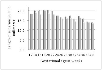
Si- Superficial inguinal ring; Ss - Scrotal sac. Positions of testes were noted according to the gestational age, weight and crown rump length (CRL) of the foetus. All testes were not descended in to inguinal canal up to the age of 24 weeks. All testes above the age of 34 weeks had reached the scrotal sac. In the period between 24 to 34 weeks testes were showing some variation in their position. In foetuses of 12 to 24 weeks (23 foetuses - 46 testes), 42 (91.3%) were Abdominal, 4 (8.7%) were at Deep Inguinal Ring. In foetuses of 26 to 34 weeks (12 foetuses – 24 testes), 2 (8.3%) were Abdominal, 3 (12.5%) were in Inguinal Canal, 5 (20.8%) were at Superficial Inguinal ring, 14 (58.3%) were in Scrotal Sac and in foetuses of 36 to 40 weeks (6 foetuses – 12 testes) all were in Scrotal sac. The gubernaculum was seen as jelly like structure extending from lower pole of testis and epididymis. Testes were sitting on gubernaculum. On all sides the gubernaculum was covered by peritoneum except on posterior aspect. The thickness of gubernaculum was more just before 24 weeks when the testes were in relation with deep inguinal ring. The dissection of inguinal region shows that the superficial and deep inguinal rings were closely approximated to each other. Inguinal canal was very small. The tip of gubernaculum was seen to be protruding outside the superficial inguinal ring. Distal attachments of ligaments of lockwood were also seen. Length of Gubernaculum was measured from lower pole of testis to the superficial inguinal ring only. Other attachments of Lockwood ligaments were ignored in these measurements. In case of full term foetus the testis were placed at the bottom of scrotal sac, in them length of scrotal ligament from lower pole of testis to base of scrotal sac was measured. The length was slightly increased from 12th that is 18.3 to 20 in 16th week then it was constant for 22nd weeks after that there was slight decrease in length every week and finally the length of scrotal ligament was 14 cm at 40th week of gestation. There was 4.3 cm decrease observed from 12th to 40th weeks (Table No. 1 and Graph No 1). Table 1: Showing the position of testis and Length of gubernaculum/Scrotal Ligament in relation with gestational age, CRL, and weight of foetuses in present study
Graph 1: Showing the Length of gubernaculum and Scrotal Ligament at different gestational ages
DISCUSSION Sampaio11 et al. reported percentage of testes in inguinal canal at different gestational ages. They found 9.5% of 10 to 23 weeks old, 58% of 24 to 26 weeks old and 21% of 27 to 30 weeks old fetuses have their testes in the inguinal canal. Sampaio11 et al. also stated that in foetuses weighing less than 500gm. (76 foetuses - 152 testes) 147 testes were in the abdomen and only 5 testes (6.5%) were in the canal. In foetuses with body weight between 501 and 1000gm. (43 foetuses – 86 testes), 54 testes (68.6%) were abdominal, 28 testes (32.5%) were in the canal and only 4 testes (4.6%) were scrotal. In foetuses with body weight between 1001 and 1500gm. (15 foetuses – 30 testes), 4 testes (13.3%) were abdominal, 3 testes (10%) canalicular and 23 testes (76.6%) scrotal. All testes were in the scrotum in foetuses weighing more than 1500 gm. (10 foetuses – 20 testes). The testes had not descended into the scrotum in any foetus weighing 740gm or less. Birnholz12 reported that the proportion of testes descending into the scrotum was 62% in the 28 to 30th weeks and 93% in the 32nd week. In present study 75% testes were descended in the 28th to 30th week in to the scrotum. Findings of these studies were comparable with the present study. C. F. Heyns8 when compared descent of testis with weight of fetus he found only one descended testis in 126 fetuses weighing less than 700 g, with the number of descended testes increasing from 22% in fetuses 700 - 999 g through 50% in those 1000-1200 g, to 72% in those with1200 g in weight. When compared with Crown Rump Length only one testis was encountered in 21 fetuses less than 209mm CRL, with descent increasing from 21 % in those of 210-239mm CRL, through 54°% in those 240-269mm CRL, to 72% in those more than 270mm CRL. Findings of this study were also comparable with the present study. The length of gubernaculum was measured from lower pole of testis to superficial inguinal ring when testes were not descended. In elderly foetuses when testes were in scrotum the length of scrotal ligament was measured from lower pole of testis to base of scrotal sac. Scrotal ligament represents the remains of gubernaculum in adult testes. The length was increased from 18.3 mm in 12th week to 20 mm in 22nd weeks after that there was slight decrease in length every week and finally the length of scrotal ligament was 14 mm at 40th week of gestation. The length was fairly constant, slightly increased prior to descent up to 22 weeks. C. F. Heyns8 also found same i.e. prior to descent of the testis there was an increase in the length of the gubernaculum as measured from the apex of the superficial inguinal ring to the inferior pole of the testis. Furthermore he also quotes that descent through the inguinal canal is a rapid process, and occurs anytime between 23 to 31 weeks of gestation. Waldemar S. Costa13 et al. in another study stated that during testicular migration gubernacular connective tissue undergoes extensive remodeling and ultimately becomes an essentially fibrous structure rich in collagen and elastic fibers. Such changes should decrease the size of the gubernaculum and, thus, contribute to other forces that cause the testes to move toward the scrotum. In fact, because of the lack of smooth muscle cells, and the amount and organization of striated muscle cells, active contraction of the gubernaculum is less likely to be an important factor in testicular descent. Both studies agree with the role of gubernaculum in descent of testes. They also agreed that length of gubernaculum remains constant during the whole gestational period. There are various theories relating to descent of testis and role of gubernaculum in it. Such as; The pulling theories propose that the contraction of striated or unstriated muscle fibres or the contracture of connective tissue intrinsic to or surrounding the gubernaculum acts to pull the testis down (Curling14 1840; Wyndham15 1943). These theories state that the gubernaculum has firm attachments both cranially and caudally, and up to 6 distal attachments have been described (Hunter3 1926) Which were named following Lockwood as ligaments of lockwood. The forces of gravity, (Hunter2 1841), peristaltic and secretory activity in the epididymis changing its centre of gravitation were also considered under pulling theories (Hadziselimovic7 1983). The findings of present study suggest that there is no active contraction of gubernaculum causing pulling of testis but length of it decreases helping in descent. The theories of pushing suggest that the testis is expelled from the abdomen by the force of increased intra-abdominal pressure, which may be the result of contraction of the abdominal wall muscles, respiratory efforts of the fetus, and the forces of labour (Hunter2 1841), distension of the bowel by meconium (Hunter3 1926), growth of the liver and other viscera (Wells4 1943) and closure of the physiological hernia (Rajfer and Walsh5 1977). These pressures are supposed to cause herniation of the gubernaculum and testis through the 'weak' part of the abdominal wall, the inguinal canal (Shrock6, 1971). The above mentioned factors have a role in descent as length of gubernaculum decreases slightly not sufficient to cause descent. Various growth theories consider testicular descent as more apparent than real, with differential growth of the lumbar vertebral column, pelvis and abdominal wall being responsible for the testis entering the inguinal canal, while the gubernaculum passively anchors it to the internal ring (Hart16 1909). Other theories have stressed the downward growth of the Processus vaginalis (Cleland17 1856); the penetrating power of unstriated muscle fibres in the gubernacular tip, enabling it to burrow its way downwards, possibly by a process of phagocytosis (Hart16 1909); dilatation of the inguinal canal and scrotum through the uptake of water by hyaluronic acid in the extracellular substance of the gubernaculum (Backhouse1 1964). Differential growth also contributes to descent as length of gubernaculum remains more less constant and also the scrotal ligament also has same length as that of gubernaculum. But the role of gubernaculum cannot be denied. The regression theories state that atrophy, degeneration or shrinking of the gubernaculum brings about testicular descent (Cleland17 1856; Hart16 1909; Rajfer and Walsh5 1977). Whatever the mechanical factors at work, there is a large body of clinical and experimental observations suggesting that the process of testicular descent is under hormonal control (Wells4 1943). Maternal, placental or fetal pituitary gonadotrophins are thought to stimulate androgen secretion by the fetal testis, with testosterone or one of its metabolites acting in some way to bring about descent of the testis (Elder et al.18 1982), also the Mullerian inhibiting substance is important for descent (Donahoe, Ito, Morikawa and Hendren19 1977). It has also been suggested that the gubernaculum is not necessary for descent at all (Wells4 1943). There is slight regression as decrease in length of gubernaculum. The values of all studies in relation with foetal weight and crown rump length were comparable with values of present study. Present study was done on aborted and stillborn normal human foetuses and no undescended testis was found in present study after 34 weeks of gestational age, foetus weighing more than 1764 gm and having Crown Rump Length more than 307 mm. The gelatinous structure of gubernaculum was changed to cord like in the late gestational period due to extensive remodeling. So the gubernaculum is important structure in descent of testis and along with several other factors helps in descent of testis. CONCLUSION In Conclusion we can state that no testis had descended to the scrotal sac up to 24th week of gestation, and all testes descended to scrotal sac at and above 34th week of gestational age. The period in which testes traversed the inguinal canal was variable from 24th to 34th week. The length of gubernaculum and scrotal ligament remained constant throughout the gestational period except a small increase in length just before the descent. The length of gubernaculum decreases slightly this decrease does not contribute to active pulling of testis hence causing descent but this helps in descent.
REFERENCES
|
|
|||||||||||||||||||||||||||||||||||||||||||||||||||||||||||||||||||||||||||||||||||||||||||||||||||||||||||||||||||||
 This work is licensed under a Creative Commons Attribution-NonCommercial 4.0 International License.