 Home
Home|
Table of Content Volume 11 Issue 3 - September 2019
A study of the diameter of femoral artery in cadavers
Dhaval K Patil1, Vaishali S Anturlikar2*
1Assistant Professor, Department of Anatomy, Seth G S Medical College, Parel, Mumbai, Maharashtra, INDIA. 2Assistant Professor, Department of Anatomy, S.M.B.T. Institute of Medical Sciences and Research Center, Dhamangaon, Tal. Igatpuri, Dist. Nashik, Maharashtra, INDIA Email: vaishali.ahire@gmail.com
Abstract Background: The femoral artery is an important artery of the lower limb from surgical and radiological point of view. Hence, the objective of this study was to measure the internal diameter of femoral artery at its origin (i.e. just below mid-inguinal point) and at its termination (i.e. at hiatus magnus). Material And Methods: The study was performed on one hundred and three lower limbs (52 of right side and 51 of left side) of properly embalmed cadavers. The various parameters of femoral artery were measured. Results: The mean internal diameter of femoral artery at its origin was 0.62 ± 0.07 cm on the right side and on the left side it was 0.63 ± 0.06 cm. The mean internal diameter of femoral artery at hiatus magnus was 0.47 ± 0.06 cm on the right side and on the left side it was 0.48 ± 0.05 cm. Conclusion: The results provide a database for the diameter of femoral artery which will be helpful for surgeons and interventional radiologists while planning and performing various clinical procedures. Key Words: Femoral artery, internal diameter, origin, hiatus magnus. INTRODUCTION The femoral artery begins as a continuation of the external iliac artery behind the inguinal ligament at the mid-inguinal point. It descends along the anteromedial part of the thigh in the femoral triangle. It then enters and courses through the adductor canal and becomes the popliteal artery as it passes through the hiatus magnus. Its first three or four centimetres are enclosed in the femoral sheath. The part of femoral artery proximal to the origin of profunda femoris artery is often termed the common femoral artery, while the part distal to the origin of the profunda femoris artery is termed the superficial femoral artery. 1Femoral artery is commonly utilized for various investigative and diagnostic procedures such as insertion of central lines, cardiac catheterization, trans arterial chemo embolization in the treatment of malignancy, arteriography in peripheral vascular diseases, angiographies as well as in magnetic resonance imaging , ultrasound and doppler imaging. The analysis of the diameter of the artery might be particularly useful in monitoring the process of the vascular remodelling in the course of atherosclerosis, hypertension, inflammation or post-radiation changes. 2
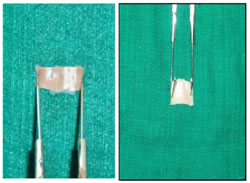
MATERIALS AND METHODS The study was done on lower limbs of fifty four embalmed cadavers (50 male and 4 female; age range: 18 - 90 years) from the department of anatomy of a tertiary care hospital. Five lower limbs were already dissected by students and hence were not included in this study. So, a total of 103 lower limbs (52 of right side and 51 of left side) were included in the study. These limbs showed no evidence of previous surgery. After taking incision, skin was reflected followed by removal of superficial fascia and deep fascia to expose the femoral triangle and adductor canal. The femoral artery was identified and exposed along its entire course. The femoral artery was cut transversely at its origin (i.e. just below mid-inguinal point MIP) and at its termination (i.e. at hiatus magnus) to measure the circumference of femoral artery as illustrated in Figure 1 and Figure 2. The internal diameter of femoral artery at its origin (i.e. just below MIP) and at its termination (i.e. at hiatus magnus) was calculated by using the formula: d = c/π (d = the internal diameter of femoral artery, c = the circumference of femoral artery). The data were statistically analysed using Microsoft Excel to calculate the mean, range and standard deviation. Figure 1 Figure 2 Figure 1: Illustration showing measurement of the circumference of femoral artery at its origin (i.e. just below inguinal ligament) Figure 2: Illustration showing measurement of the circumference of femoral artery at its termination (ie. at hiatus magnus) RESULTS The mean internal diameter of femoral artery at its origin was 0.62 ± 0.07 cm on the right side and on the left side it was 0.63 ± 0.06 cm. The range of internal diameter of femoral artery at its origin was 0.41 – 0.76 cm in 52 right lower limbs and 0.48 – 0.83 cm in 51 left lower limbs (Table 1). TABLE 1: Table showing the distribution of limbs with respect to the internal diameter of femoral artery at its origin
The mean internal diameter of femoral artery at hiatus magnus was 0.47 ± 0.06 cm on the right side and on the left side it was 0.48 ± 0.05 cm. The range of internal diameter of femoral artery at hiatus magnus was 0.35 – 0.61 cm in 52 right lower limbs and 0.37 – 0.61 cm in 51 left lower limbs (Table 2). TABLE 2: Table showing the distribution of limbs with respect to the internal diameter of femoral artery at its termination
DISCUSSION So far, femoral artery diameters have been studied by various authors by various methods for few segments of the artery in smaller clinical samples.The study by Horejs D et al 3 in the year 1988 of 130 males revealed mean diameters of 1.05 cm and 1.04 cm for the right and left common femoral arteries, respectively in the age group of 50–60 years using computed tomography.In 1999, Sandgren T4 et al measured the diameter of the femoral artery in 122 healthy volunteers with ultrasound scan and found that the mean diameter of the femoral artery were 9.8 mm in males and 8.2 mm in females. In 2000, study by G. Rådegran et al 5 found that the diameter of common femoral artery determined by ultrasound Doppler was 10.6 ± 0.4 mm (range 8.2–12.7 mm)In 2000, P Hughes et al 6 studied the femoral artery diameter in fifty patients using a portable ultrasound machine. They found the diameter of the femoral artery as 0.9 ± 0.3 cm (Range: 0.5-2.9); 0.8± 0.2 cm (Range: 0.4-1.3) and 0.7 mm ± 0.1 cm (Range: 0.4-1.2) at the level of inguinal ligament; 2 cm below the inguinal ligament and 4 cm below the inguinal ligament respectively. In 2001, Kenneth Spector et al 7 analysed 60 consecutive peripheral angiograms which revealed an average vessel diameters of 6.6 mm (Range: 3.9–8.9 mm) and 5.2 mm (Range: 2.5–9.6 mm) for the common femoral artery and superficial femoral artery, respectively. In 2007, Baptist M et al 8, dissected 40 lower limbs and found that the internal diameter of the femoral artery at its origin ranged between 0.6 - 1.0 cm. In the 2012 study by Dorota Czyżewska et al 2 using ultrasound assessment in 228 healthy persons in Poland found that the mean diameter of femoral artery 3 cm above the origin of profunda femoris artery was 8.05 ± 1.07 mm. The mean diameter of femoral artery 3 cm below the origin of profunda femoris artery was 6.06 ± 0.69 mm. In 2013, Shiny Vinila B H et al 9 dissected 40 lower limbs and found that the average internal diameter of femoral artery was 7.02 ± 0.85 mm. In 2016, study by Swetha B et al 10 on 20 embalmed cadavers, the average internal diameter of the femoral artery was 8.5 mm. In 2018, a study by K Rajeswari et al 11 measured the diameter of femoral artery at the level 1 cm. below the inguinal ligament by three different methods. The diameter of the femoral artery observed was 7.7mm (Range: 7 - 9.5 mm); 7.5 mm (Range: 6.8 - 8.2 mm) and 7.6 mm (Range: 6.8 - 11 mm) by direct dissection method; by silicone gel mould method and by observation of the 64 slice computerised tomographs respectively.In the 2018 study by Roberto Lorbeer et al 12, Gadolinium-enhanced magnetic resonance angiography at 1.5 Tesla was performed in 756 male participants. The mean diameter of proximal femoral artery on left side was 0.812 ± 0.120 cm and on right side was 0.803 ± 0.117 cm. The mean diameter of distal femoral artery on left side was 0.593 ± 0.093 cm and on right side was 0.594 ± 0.091 cm.In the present study, the mean internal diameter of femoral artery at its origin was 0.62 ± 0.07 cm on the right side and on the left side it was 0.63 ± 0.06 cm. The mean internal diameter of femoral artery at hiatus magnus was 0.47 ± 0.06 cm on the right side and on the left side it was 0.48 ± 0.05 cm.The differences in the diameter of femoral artery measured by various studies are due to differences in methods, techniques and sites used for measurement as well as racial and ethnic variations.
CONCLUSION The knowledge of anatomy of the femoral artery is crucial from point of view of surgeons, interventional radiologist and physicians. So the diameter of femoral artery at the origin and termination were measured and compared with available literature. This comparison will be useful for clinicians in performing various procedures on femoral artery and in reducing the chances of procedural complications.
REFERENCES
|
|
||||||||||||||||||||||||||||||||||||||
 This work is licensed under a Creative Commons Attribution-NonCommercial 4.0 International License.