 Home
Home|
Table of Content Volume 11 Issue 3 - September 2019
A study of morphometry of posterior Talo-fibular ligament in the talocrural joint
Shishirkumar1, Shivarama C H2*, Roshan S3, ChethanaY K4, Nishitha5
1,2Assistant Professor, 3Associate Professor, 4,5Tutor Department of Anatomy, Kanachur Institute of Medical Sciences, Mangalore Email: drshivac@gmail.com
Abstract Background: Population under study is habitual squatters and an undue stretch is applied on the posterior-talo-fibular ligament in doing so. This study is done to find the morphometry of the posterior talo-fibular ligament as it is not done in this population. The population under study is as discussed are habitual squatters and an enquiry is done so as to find out whether any morphometric difference is there when compared to the other literature. Materials and Methods: Thirty formalin fixed cadavers are dissected. All thirty are dissected in the Department of Anatomy, Kanachur Institute of Medical Sciences, Mangalore. Results: The thickness difference between males and females is statistically significant(p=0.041). Conclusion: This study can be considered as a reference point and further studies has to be carried out pan India to know the difference. Key Words: Squatting, Morphometry, Talo-Crural Joint, Talo-fibular Ligament.
INTRODUCTION Posterior Talo-Fibular Ligament is a very strong ligament and has a near horizontal position. The ligament originates on the medial surface of the lateral malleolus from the lower segment of the digital fossa. It courses horizontally and is inserted on lateral surface of the talus in a groove along the posteroinferior border of the lateral malleolar upto its midsegment and also to the posterior surface of talus. Indians are known to be habitual squatters1,2. Chimba Mkandawire, et al3, in 2005 in their study on the foot and ankle ligament morphometry, using 121 bone-ligament preparations from 26 cadaver feet, the following measurements were noted, Posterior talofibular mean length was measured to be 27.74 ± 3.41 mm. Mahmut Ugurlu et al4. in 2010 studied the anatomy of the lateral complex of the ankle joint in relation to peroneal tendons, distal fibula and talus in 22 formalin fixed ankles. In their study, the posterior talo-fibular mean length was measured to be 24.12 mm and the mean width of 5.09 mm. Muzaffer Sindel et al5. in 1998 on their study on the lateral ankle ligaments by in 24 ankles, mentioned that the posterior talo-fibular ligament, the mean length was 20.7 mm with a standard deviation of 2.15 mm; the mean width was 6.1 mm with a standard deviation of 0.77 mm. This study is done to find the morphometry of the posterior talo-fibular ligament as it is not done in this population. The population under study are habitual squatters and an enquiry is done so as to find out whether any morphometric difference is there when compared to the other literature.
AIMS AND OBJECTIVES To Study the Morphometry of Posterior Talo-Crural Joint.
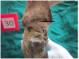
MATERIALS AND METHODS Thirty formalin fixed human ankles were dissected which was available in the department of anatomy, Kanachur Institute of Medical Sciences, Mangalore in 15 cadavers. Male and female ankles were categorized and also right from the left. The study was done from September 2016 to August 2018. Incision was made on the anterior median plane and posterior median plane from caudal one third of leg to proximal one third of foot. Skin was reflected all around the talocrural joint till the meeting of dorsal surface and plantar surface. All the soft tissues including the muscles were dissected and reflected on the anterior, posterior, medial and lateral surfaces. The soft tissue tunnel which surrounds the tendons of muscles is in intimate relation with the underlying ligaments of the talocrural joint. The posterior talofibular ligament was exposed and since it is a cord like ligament only the length and breadth measurements were taken and thickness was measured. Table 1: Morphometry of the Posterior Talo-Fibular Ligament
Note:R-Right L-Left M-Male F-Female
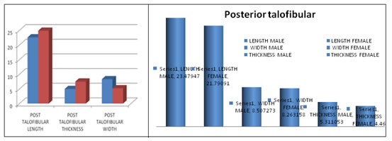
Graph 1: Right Vs Left Graph 2: Male Vs Female
Irrespective of the side and sex to which the ligaments belong the mean value of the length of the posterior talo-fibular ligament is 22.86 mm. The widths in is 8.35 mm .The thickness mean measurement is 4.99 mm The mean length value on the right side is 23.14 mm. The mean width value is 8.41 mm. The mean thickness measurement is 5.03 mm. The mean length value on the left side is 22.57 mm. The mean width value is 8.29 mm. The mean thickness measurement is 4.96 mm .The mean length value in male is 23.47 mm. The mean width value is 8.50 mm. The mean thickness measurement is 5.3 mm. The mean length value in female is 21.79 mm. The mean width value is 8.26 mm. The mean thickness measurement is 4.46 mm. The thickness difference between males and females is statistically significant(p=0.041). Table 2: Variations in talofibular ligament the posterior
Table 3:
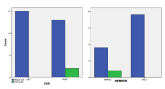
Graph 3: (left) indicates side and graph no. 4(right) indicates sex Variations found in the posterior tibiofibular ligament in the form of slip to the tibia in two cases out of thirty accounting to 6.66% of the total. It is found in 13.3% on the right side, 18.2% in females.
DISCUSSION Irrespective of the side and sex to which the ligaments belong the mean value of the length of the posterior talo-fibular ligament is 22.86 mm. The widths in is 8.35 mm .The thickness mean measurement is 4.99 mm. It is a cord like ligament. The mean length value on the right side is 23.14 mm with a standard deviation of 3.55 mm. The mean width value is 8.41 mm with a standard deviation of 1.45 mm. The mean thickness measurement is 5.03 mm with a standard deviation of 1.22 mm. The mean length value on the left side is 22.57 mm with a standard deviation of 2.68 mm. The mean width value is 8.29 mm with a standard deviation of 1.15 mm. The mean thickness measurement is 4.96 mm with a standard deviation of 1.02 mm The right and the left side measurements are almost symmetrical on both sides. The mean length value in male is 23.47 mm with a standard deviation of 2.93 mm. The mean width value is 8.50 mm with a standard deviation of 1.53 mm. The mean thickness measurement is 5.31 mm with a standard deviation of 1.00 mm The mean length value in female is 21.79 mm with a standard deviation of 3.24 mm. The mean width value is 8.26 mm with a standard deviation of 1.16 mm. The mean thickness measurement is 4.46 mm with a standard deviation of 1.13 mm In males the measurements are consistently higher than that of females. The thickness difference between males and females is statistically significant. This may account for more lateral ligaments sprain in females when compared to the males. According to Milner and Soames6, on the anatomy of collateral ligaments of human ankle joint (1998), the mean length was 23 mm, with a standard deviation of 7 mm and the mean width was measured to be 5.5 mm with a standard deviation of 2.5 mm. According to Taser and Co7, the mean length was measured to be 21.66 mm with a standard deviation of 4.8 mm and mean width was 5.55 mm with a standard deviation of 1.3 mm On the anatomy of lateral ankle ligaments by Muzaffer and Sindel and Co6, the posterior talo-fibular ligament, the mean length was 20.7 mm with a standard deviation of 2.15 mm; the mean width was 6.1 mm with a standard deviation of 0.77 mm Chimba Mkandawire et al3 (2005) in their study on “The Foot and ankle ligament morphometry.” in 121 bone- ligament- bone preparations from 26 cadaver feet. Posterior talofibular mean length was measured to be 27.74 ± 3.41 mm.On a study of anatomy of lateral complex of the ankle joint in relation to peroneal tendons, on the distal fibula and talus by Mahmut Ugurulu4, the posterior talo-fibular mean length was measured to be 24.12 mm and the mean width of 5.09 mm The measurements in other studies are congruent with our study. Variations in the posterior talofibular ligament In our study the variations was found in the posterior tibiofibular ligament in 6.66% of the total. It is found in 13.3% on the right side. It is found in 18.2% in females. The inter malleolar ligament (a part of posterior talo-fibular ligament) was found in 81.8 percent of the specimen, according to Pau Galeno, in his study of anatomy of ankle ligaments The non congruency may be due to the population studied in our study is different from the other studies.
CONCLUSION This study can be considered as a reference point and further studies has to be carried out pan India to know the difference.
REFERENCES
|
|
||||||||||||||||||||||||||||||||||||||||||||||||||||||||||||||||||||||||||||||||||||||||||||||||||||||||||||||||||||||||||||||||
 This work is licensed under a Creative Commons Attribution-NonCommercial 4.0 International License.