 Home
Home|
Table of Content Volume 11 Issue 3 - September 2019
A study of morphometry of calcaneo-fibular ligament in the Talo-crural joint
Shishirkumar1, Shivarama C H2*, Roshan S3, Chethana Y K4, Nishitha5
1,2Assistant Professor, 3Associate Professor, 4,5Tutor, Department of Anatomy, Kanachur Institute of Medical Sciences, Mangalore. Email: drshivac@gmail.com
Abstract Background: Habitual squatting has long been recognised to alter the skeletal morphology of the lower limb. Squatting is a resting postural complex that involves hyperexion at the hip and knee and hyperdorsiflexion at the ankle and subtalar joints. During locomotion, the foot is rarely dorsifexed sufficiently to bring the anterior border of the inferior extremity of the tibia into contact with the dorsum of the neck of the talus. Thus modifcations of any type in the ligaments indicating their habitual contact have been taken as evidence of the extreme dorsiflexion of the ankle that occurs in squatting is the question to be asked. This study pputs in a sincere effort to find the morphometry of calcaneofibular Ligament. Materials And Methods: Thirty formalin fixed cadavers are dissected. All thirty are dissected in the Department of Anatomy, Kanachur Institute of Medical Sciences, Mangalore. Results: The study is in agreement with the values of that was found in other studies which it was compared. Conclusion: This study can be considered as a reference point and further studies has to be carried out pan India to know the difference. Key Words: Squatting, Morphometry, Talo-Crural Joint, Calcaneofibular Ligament. INTRODUCTION Calcaneofibular ligament is a strong, cordlike or flat oval ligament. It originates from the lower segment from the anterior border of the lateral malleolus. In neutral position of the foot it runs posteriorly, inferiorly and medially. It is inserted on a small tubercle located on the posterior aspect of the lateral calcaneal surface. Chimba Mkandawire, et al1, in 2005 in their study on the foot and ankle ligament morphometry, using 121 bone-ligament preparations from 26 cadaver feet, the following measurements were noted. Calcaneofibular mean length was measured to be 35.44 ± 6.31 mm. Mahmut Ugurlu et al2. in 2010 studied the anatomy of the lateral complex of the ankle joint in relation to peroneal tendons, distal fibula and talus in 22 formalin fixed ankles. In their study, the calcaneo-fibular ligament measured a mean length of 26.67 mm and a mean width of 4.57 mm. Muzaffer Sindel et al3. in 1998 on their study on the lateral ankle ligaments by in 24 ankles, mentioned that the calcaneo-fibular ligament measured a mean length of 26.8 mm and a mean width of 6mm. Kitsoulis .P. et al4. in 2011 in their morphological study of the calcaneofibular ligament in 72 embalmed human ankles determined the anatomy of the calcaneofibular ligament. The calcaneofibular ligament originates from the lower segment of the anterior border of the lateral malleolus and after a posterior, inferior, and medial course inserts on a small tubercle of the lateral calcaneal surface. The ligament is extracapsular and cord-like. There were 52 subjects presenting one band forming the CFL, 16 with two bands and 4 with three bands. We recorded the presence of 24 ankles where the CFL had a common origin with the anterior talofibular one. There were also 4 cases where the anterior TFL was absent. The mean measured length of the CFL was 31.83 mm, the mean width 4.42 mm, and the thickness was 1.58 mm on average. The mean angle formed in relation to the sagittal plane was 52.11°. The maximal elongation of the CFL during pronation was 2.88 mm on average concerning both sexes. Women presented 3.28 mm elongation of CFL on average during maximum inversion and dorsiflexion while in men it was 2.63 mm. This study puts in a sincere effort to find the morphometry of the ligament in our population since we are geographically and ethinically so different. AIMS AND OBJECTIVES To study the morphometry of the calcaneo fibular ligament in our population.
MATERIALS AND METHODS Thirty formalin fixed human ankles were dissected which was available in the department of anatomy, Kanachur Institute of Medical Sciences, Mangalore in 15 cadavers. Male and female ankles were categorized and also right from the left. The study was done from September 2016 to August 2018. Incision was made on the anterior median plane and posterior median plane from caudal one third of leg to proximal one third of foot. Skin was reflected all around the talocrural joint till the meeting of dorsal surface and plantar surface. All the soft tissues including the muscles were dissected and reflected on the anterior, posterior, medial and lateral surfaces. The soft tissue tunnel which surrounds the tendons of muscles is in intimate relation with the underlying ligaments of the talocrural joint. The calcaneofibular ligament was exposed and since it is a cord like ligament only the length and breadth measurements were taken and thickness was measured.
RESULTS Table 1: Morphometry of Calcaneo-Fibular Ligament
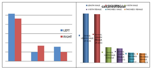
Graph 1: Morphometry of Calcaneo-Fibular Ligament Graph 2: Male Vs Female
Irrespective of the side and sex to which the ligaments belong the mean value of the length of the calcaneo-fibular ligament is 25.30 mm. The widths in is 7.66 mm. The thickness mean measurement is 5.09 mm. The mean length value on the right side is 24.82 mm. The mean width value is 7.49 mm. The mean thickness measurement is 5.19 mm. The mean length value on the left side is 25.78 mm. The mean width value is 7.83 mm. The mean thickness measurement is 4.99 mm. The mean length value in male is 25.51 mm. The mean width value is 8.08 mm. The mean thickness measurement is 5.21 mm. The mean length value in female is 25.18 mm. The mean width value is 7.42 mm. The mean thickness measurement is 4.87 mm. DISCUSSION Calcaneofibular ligament Irrespective of the side and sex to which the ligaments belong the mean value of the length of the calcaneo-fibular ligament is 25.30 mm. The widths in is 7.66 mm. The thickness mean measurement is 5.09 mm. It is a cord like ligament. The mean length value on the right side is 24.82 mm with a standard deviation of 2.42 mm. The mean width value is 7.49 mm with a standard deviation of 1.36 mm. The mean thickness measurement is 5.19 mm with a standard deviation of 1.44 mm The mean length value on the left side is 25.78 mm with a standard deviation of 2.40 mm. The mean width value is 7.83 mm with a standard deviation of 1.19 mm. The mean thickness measurement is 4.99 mm with a standard deviation of 1.11 mm The right and the left side measurements are almost symmetrical on both sides. The mean length value in male is 25.51 mm with a standard deviation of 2.30 mm. The mean width value is 8.08 mm with a standard deviation of 1.12 mm. The mean thickness measurement is 5.21 mm with a standard deviation of 0.92 mm The mean length value in female is 25.18 mm with a standard deviation of 2.54 mm. The mean width value is 7.42 mm with a standard deviation of 1.31 mm. The mean thickness measurement is 4.87 mm with a standard deviation of 1.75 mm In males the measurements are consistently higher than that of females. It may be because the males are well built than females. Chimba Mkandawire et al1 (2005) in their study on “The Foot and ankle ligament morphometry.” in 121 bone- ligament- bone preparations from 26 cadaver feet. Calcaneofibular mean length was measured to be 35.44 ± 6.31 mm According to the study by Ruth CJE et al5, the length ranged from 30 mm to 40 mm and width ranged from 4 mm to 5 mm According to Prins J G et al6, on the diagnosis and treatment of injury to the lateral ligament of ankle, the mean length measured 20mm, the mean width measured 5mm and mean thickness measured 3mm According to Mahmut Ugurlu2, on the anatomy of lateral complex of the ankle joint in relation to peroneal tendons, distal fibula and talus, the calcaneo-fibular ligament measured a mean length of 26.67 mm and a mean width of 4.57 mm According to Milner and Soames7, the mean length measured 19.5 mm with a standard deviation of 3.9 mm; the mean width measured 5.5 mm with a standard deviation of 1.6 mm According to Taser et al8, the mean length measured 31.94 mm with a standard deviation of 3.7 mm and the mean width measured 4.68 mm with a standard deviation of 1.3 mm According to P.Kitsoulis et al4, on morphological study of calcaneo-fibular ligament of in 72 cadaveric lower limbs, the mean length is 31.83 mm, the mean width was 4.42 mm and the mean thickness was 1.58 mm According to Muzaffer and Sindel et al3, on the anatomy of lateral ankle ligaments, the calcaneo-fibular ligament measured a mean length of 26.8 mm and a mean width of 6 mm The measurements in the studies are congruent with our study measurements.
CONCLUSION The study has been successful in finding out the morphometry of this ligament. The measurements in the other studies are congruent with our study measurements. We are in strong agreement when compared to the other studies.
REFERENCES
|
|
|||||||||||||||||||||||||||||||||||||||||||||||||||||||||||||||||||||||
 This work is licensed under a Creative Commons Attribution-NonCommercial 4.0 International License.