 Home
Home|
Table of Content Volume 12 Issue 1 - October 2019
A study on variations of the musculocutaneous nerve in relation to the coracobrachialis muscle
J Gayathri1, M Bharatha Devi2*, V Anandhi3
1Assistant Professor, Department of Anatomy, Thanjavur Medical College, Thanjavur, Tamilnadu , INDIA. 2Assistant Professor, 3Professor and HOD Department of Anatomy, K A P V Government Medical College, Trichy-1, Tamilnadu, INDIA. Email: drbharathigandhi@gmail.com
Abstract Background: The brachial plexus is formed by the union of the ventral rami of C5-T1 spinal nerves. Contributions to the plexus by C4 and T2 differ. The musculocutaneous nerve (MCN) originates from the lateral cord of brachial plexus and supplies the coracobrachialis, biceps brachii and brachialis muscles in the anterior compartment of arm. Then it continues as the lateral cutaneous nerve of the forearm. Variations in the origin, course, branching pattern, termination and the connections of the musculocutaneous nerve are not uncommon. Methods: 25 embalmed cadavers were taken for the study during the period of 2 years in the Department of Anatomy, K.A.P.V Government Medical College,Trichy. The dissection was done on the pectoral region, axilla and the arm. The infraclavicular part of the brachial plexus was exposed. The musculocutaneous nerve was identified. The origin, number, course and its relation to the coracobrachialis muscle were studied. Results: In this study, in 12% of specimens, the musculocutaneous nerve (MCN) had 2 branches. The unnamed medial branch of musculocutaneous nerve (MCN) communicated with the median nerve without piercing the coracobrachialis muscle in 2% of specimens and after piercing the coracobrachialis muscle in 4% of specimens. The nerve did not pierce the coracobrachialis in 6% of the upper limbs. Conclusions: Knowledge of variations of musculocutaneous nerve (MCN) is essential during surgical procedures involving upper limb, in brachial plexus blocks, in post traumatic evaluations and in exploring the arm for peripheral nerve repair. Key Words: brachial plexus, coracobrachialis, median nerve, musculocutaneous nerve, unnamed nerve,)
INTRODUCTION The brachial plexus is formed by the union of the ventral rami of C5 – T1 nerve roots. It has root, trunk, divisions, cords and branches. The cords have 3 subdivisions namely medial, lateral and posterior. The musculocutaneous nerve (MCN) originates from the lateral cord of brachial plexus and supplies the coracobrachialis,1,2 biceps brachii and brachialis muscles in the anterior compartment of the arm and then continues as the lateral cutaneous nerve of the forearm3. The median nerve is formed by medial and lateral roots derived from medial and lateral cords. The coracobrachialis is located in superomedial part of the arm and it is pierced by MCN, the distal part of its attachment indicates the location of the nutrient foramen of humerus. Variations in the formation of the brachial plexus and its terminal branches in the upper arm are not uncommon and have been reported previously. The anatomical variations of the musculocutaneous nerve4 and the median nerve are related to embryological development. The course and branching anomalies of the musculocutaneous nerve and its relation to the coracobrachialis muscle have been described by Koizumi1, Buch5,Flatow et al6 and Le Minor7.
MATERIALS AND METHOD 50 upper limbs from 25 embalmed cadavers were taken for the study during the period of 2 years during routine dissection in the Department of Anatomy, K.A.P.V Government Medical College, Trichy. The dissection was performed according to the standard procedures of Cunningham’s Manual of Practical Anatomy Vol I. RESULTS Specimen No 1 The following variation was found in the right upper arm. The right median nerve was formed by lateral and medial roots as usual. Lateral to the lateral root of median nerve, the musculocutaneous nerve (MCN) was split into two at the level of formation of the median nerve (fig-1). The lateral part of MCN pierced the coracobrachialis muscle in the middle part and after supplying biceps and brachialis, continued as the lateral cutaneous nerve of forearm. Proximal to the coracobrachialis muscle, the medial part of MCN joined the median nerve without piercing the coracobrachialis muscle and it did not supply any other muscle in the arm.
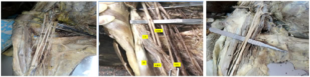
Figure 1: (Type 1: The communication is Figure 2: (Type 2: The communication is Figure 3: (Type 3: The branches and the nerve Proximal to coracobrachialis muscle) distal to coracobrachialis muscle) itself did not pierce the coracobrachialis) CB:Coracobrachialis, BB:Biceps brachi,MN:Median nerve, MCN: Musculocutaneous nerve,UN: Ulnar nerve ,LR:Lateral root of median nerve,MR:Median root of median nerve. CB:Coracobrachialis, BB:Biceps brachi, MN:Median nerve, MCN: Musculocutaneous nerve CB:Coracobrachialis, BB:Bicepsbrachi, MN:Median nerve ,MCN:Musculocutaneous nerve, LCNA: Lateral cutaneous nerve of arm.
Specimen no: 2 The above variation was observed in the right upper arm. The right median nerve was formed by lateral and medial roots as usual. Lateral to the lateral root of median nerve, the MCN split into two after travelling a short distance while parallel to the median nerve. Both the nerves pierced the coracobrachialis muscle in the middle part (fig-2).The lateral branch of MCN continued as the lateral cutaneous nerve of the forearm. The medial part of the MCN,after piercing the coracobrachialis muscle rejoined the median nerve and it did not supply any other muscle in the arm. The communication was found distal to coracobrachialis muscle. Third variation: In three specimens, the MCN did not pierce the coracobrachialis muscle(fig-4), it continued as lateral cutaneous nerve of forearm passing between brachialis and biceps muscle. There was no communication noted between median nerve and musculocutaneous nerve.
DISCUSSION During embryogenesis, each myotome and dermatome maintains its own innervations of the upper limb formation. During the 5th week of intrauterine life, the muscles are developed from the mesenchymal cells of paraxial mesoderm by the expression of 5 HOX D genes. The spinal nerve axons grow distally and reaching the mesenchyme of the developing limb. During this process, some of the nerves form tight connections to one another and subsequently join into specific variations of the brachial plexus8. Various authors reported communicating branches between the MCN and median nerve at different levels 9,10,11. Five types of variation were reported7. Type I: The MCN pierces the coracobrachialis muscle and innervates all the anterior compartment muscles of arm. There are no connecting fibers between the musculocutaneous and median nerve12,13 Type II: The main trunk of median nerve is formed by union of few fibers of the lateral and median root of the median nerve . The remaining medial root fibers pass in the MCN leaving it after a distance to join the main trunk of median nerve. Type III: The lateral root of the median nerve passes in the MCN and leaves it after a distance to join the main trunk of median nerve. Type IV: The MCN fibres unite with the lateral root of the median nerve and emerge from median nerve at various level in arm in a short distance. Type V: The MCN is absent. The fibers of MCN run within the median nerve along its course. In this type, the MCN does not pierces the coracobrachialis muscle, the variation of Type V was described by Broca in 1888. According to Le Minor, incidence of MCN variation was about 0.3-2%7. The variation found in only 3 cases (1.7%) among 175 brachial plexuses studied by Kerr14. Watanabe et al found the communication between musculocutaneous and median nerve only in 2 cases (1.4%) among 140 upper limbs15. Here, the present study correlates with Type II and Type III ,the unnamed branch of MCN united with the lateral root of the median nerve, and rejoined the median nerve. The MCN has rather constant anatomical features, originating from the lateral cord of the brachial plexus and piercing the coracobrachialis muscle. However, Buch5 reported in his cadaveric study, that the MCN originated from the median nerve in 3-6% and from the posterior cord in 1-5% of specimens. Le Minor7, Spinner and Winkelman16 observed that the lateral cord, without giving off the lateral root of the median nerve, passed through the coracobrachialis muscle and innervated the coracobrachialis, biceps brachii and brachialis muscles. According to Buch, up to 14%, the MCN did not pierce the coracobrachialis muscle or might be absent in some cases1. In the present study, the MCN did not pierce the coracobrachialis in 3 specimens. Venierators and Anangnostopoulou 9,10 described three different types of communication between the musculocutaneous nerve and median nerve in relation to the coracobrachialis muscle. Type 1: The communication is proximal to coracobrachialis muscle. Type 2: The communication is distal to coracobrachialis muscle. Type 3: The MCN and its branches are not seen to pierce the coracobrachialis. There are many reports of the occurrence of a communication between the musculocutaneous nerve and the median nerve5,15. In one case, the MCN was communicated with the median nerve after piercing the coracobrachialis, which was reported by Joshi17 and in 3.125% of the cases reported by Bhattarai4. In few cases, instead of the whole trunk of the nerve piercing the coracobrachialis, only its muscular branch was found to pierce the muscle. Instead of piercing the coracobrachialis, the nerve may pass behind it or between it and the short head of the biceps muscle. Occasionally, the nerve penetrates not only the coracobrachialis, but also the short head of the biceps muscle or the brachialis. In the present study, there is communication between median nerve and the unnamed medial branch of MCN, proximal to the coracobrachialis muscle in 2%. In 2 specimens the MCN were found to rejoin the median nerve after piercing the coracobrachialis muscle. This communication is distal to the coracobrachialis muscle. The musculocutaneous nerve did not pierce the coracobrachialis in 3 specimens. During embryonic life, some factors influencing the mechanism of formation of limb muscles and peripheral nerves which leads to anatomical variations of MCN.
CONCLUSION The variations of MCN have significance during surgical procedures, in the brachial plexus block, in post traumatic evaluations and in the exploratory innervations of the arm for peripheral nerve repair. Awareness and knowledge of these variations is necessary for the surgical managements and intervention procedures in radiology.
REFERENCES
|
|
 This work is licensed under a Creative Commons Attribution-NonCommercial 4.0 International License.