 Home
Home
|
Table of Content Volume 12 Issue 2 - November 2019
Correlative study of diameter of carotid canals with cranial index in Gujarat population
Aakruti Rasiklal Parmar1*, Yogesh N Chauhan2
1Associate professor, Department of Anatomy Government Medical College Bhavanagar-364001, Gujarat, INDIA. 2Consultant Obstestrics and Gynaecology, Chiranjivi Hospital, Sihor, Gujarat, INDIA. Email: aakrutiparmar@yhoo.com
Abstract Background- 42 Male and 28 female non- pathological dried crania were studied to correlate the CI of both male and female crania with diameters of both right and left carotid canals. It will help to predict or explore the carotid canals by knowing the CI of crania and vice versa. Method- Cranial Index was measured by winged caliper. Carotid canals by digital venire caliper. The obtained results were correlated with coefficient regression equations Results-The correlation between CI and diameter of Rt CC 0.06 and P value 0.712 CI and diameter of Lt CC correlative value (r value) .0.15 P value 0.352. In female crania correlation between CI and Diameter of Rt CC was -0.13 and P value was 0.513, correlation between CI and Diameter of CC was-0.30 and P value was 0.129. The regression analysis of CI as dependent variable and Rt CC and Lt CC as predictors–constant regression coefficient was 72.9 (SE 2.0) t=35.2 and (P<0.01) highly significant, Diameter of Rt CC had regression coefficient was- 1.26 (SE 2.4) t= 0.507, P=0.61. Diameter of left CC regression coffe-2.14 (SE-2.4) t= 0.89 P=0.37, Male compared to female regression coefficient was-4.47(SE-0.911) t= 4.90 (P<0.01) Conclusion- This correlative coefficient and regression equation of CI with Rt. and Lt. CC will be quite helpful to Neurosurgeon, Neuron physician, anatomist, anthro pologist, medico-legal expert Key Word: CI= cranial Index, CC= carotid canal, correlative coefficient, Regression, winged caliper, vernier caliper
INTRODUCTION The carotid canal or canals caroticus (CC) is a large, almost circular foramen, through the petrous-part of temporal bone. It transmits internal carotid Artery (ICA) with venous plexus and some autonomic nerves, CC found postero-lateral to the foramen lacerum and lateral to the Jugular foramen1,2. The opening of CC is found in front foramen lacerum and ICA enters the cranial cavity intracranially. The ICA separated from CC walls by the peri-vascular tissues and venous plexus3. Basilar cranial fracture has been associated with injury to ICA and vascular complications are more frequently observed when there was involvement of CC. Fracture through petrous segment also involves the carotid canal is associated with a relatively high incidence of carotid injury and patients suffer more severe head injury followed by haemorrage4. To know the degree of fracture and extent of involvement of ICA it is mandatory to know the diameter of CC on both sides. It was very difficult to visualize radiologically due to severe bleeding. Hence attempt was made to study the diameters of both and right and left CC and correlated with CI of the same crania so-that by knowing the CI of the patients Neuro-physician or surgeon can predict the diameter of CC Moreover By knowing the diameter CC one can explore the CI of the individual which has great importance in medico- legal practice.
MATERIAL AND METHODS 42 male and 28 female, crania available in the anatomy and forensic department of Government medical college Bhavnagar- 364001 Gujarat were studied. Inclusive criteria- Non- pathological, dried crania, after confirmation of gender differences, adult crania were selected. Exclusion criteria – Broken, Pathological, crania were excluded from the study. Method- Each crania was put in a anatomical position. Cranial Index was measured from Nasion to Inion and one supramastoid crest to another supra- mastoid crest and divided by 100. CI= Maximum breadth of crania X100 Maximum length crania The diameters of carotid canal were measured by digital venire caliper. The obtained results were correlated. The duration of study was April 2005 to October 2019 Statistical analysis- The obtained results were correlated coefficiently and regression values were also noted. The statistical analysis was performed in SPSS 2007 software.
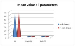
OBSERVATION AND RESULTS In male crania mean value of CI was 65.16 (SD±5.70) and females CI was 68.9(SD±4.20) and (P<0.01) P value was significant. Table 1: Mean value all parameters
The mean value of Rt CC in Male crania was 0.9.1 (SD±0.1) and P value was highly significant (P<0.01) The mean value of Lt CC female crania was 0.60(SD±0.1) and female was 0.40 (SD±1) and P value was significant (P<0.01)
Table 2: Pearson Correlation coefficient between cranial Index and Diameter of carotid canals
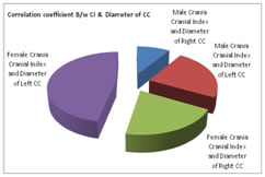
Correlation coefficient between Index and dimensions of right and left carotid canals. In male crania – correlation between CI and right diameter of right carotid canal (Rt CC) was 0.06 and P value was 0.71, CI with diameter with left carotid canal was (it CC) was 0.15 and P value was 0.35. In female crania- correlation coefficient between CI with Rt CC was -0.13 and P value was 0.51 CI with Lt CC was 0.30 and ‘t’ value was 0.12 and P value was highly significant (P<0.01)
Table 3: Regression Analysis of Cranial index as depended variable and other predictors
Note* significant at 5% level of significance (P<0.05) Adjusted R Square 0.829
Table 3: In the regression analysis as CI was depended variable and Rt and it CC were predictors. The regression coefficient was 72-9 (SE-2.07) ‘t’ value was 35.2 and P value was highly significant (P<0.01). In the diameter of Rt CC regression coefficient was- 1.26(SE-2.4) t= -0.50 and P value was 0.614. Diameter of lt CC. regression coefficient was -2.14(SE-2.4)t=-0.89 and P value =0.37. Male (Compared to female) regression coefficient was -4.471(SE-0.91) t=- 4.90 and P <0.01 (highly significant)
DISCUSSION In the present correlative study of diameter of CC with CI in Gujarat population-The mean value of male CI was 65.16 (SD±5.7) and females CI 68.9(SD±4.20) and (P<0.01). The mean value of Rt CC in Male crania was 0.9.1 (SD±0.1) and female crania was 0.60(SD±0.1) and female was 0.40 (SD±1) and P value was significant (P<0.01) (Table-1) The Pearson Correlation coefficient between CI and dimensions of right and left carotid canals. In male crania – correction between CI and right diameter of right carotid canal (Rt CC) was 0.06 and P value was 0.71, CI with diameter with left carotid canal was (Lt CC) was 0.15 and P value was 0.35. In female crania – Correlation coefficient between CI and Rt CC was -0.13 and P= 5.13. Correlation between CI and Lt. CC was-0.30 P=0.129(Table-2). In the regression analysis as CI was depended variable and Rt and Lt CC were predictors. The regression coefficient was 72-9 (SE-2.07) ‘t’ value was 35.2 and P value was highly significant (P<0.01). In the diameter of Rt CC regression coefficient was- -126(SE-2.48) t= -0.50 and P value was 0.614. Diameter of Lt CC. regression coefficient was -2.14(SE-2.4) t=-0.89 and P value =0.3. Male (Compared to female) regression coefficient was -4.471(SE-0.91) t=- 4.90 and P <0.01 (highly significant) (Table-3). These findings were more or less in agreement with previous studies5,6,7.It is interesting to note that CI of female is more due to more width of female crania but diameters of CC were lesser than male crania. The probable reason could be, hemodynamic pressure is more in males than females hence diameters of female CC remain smaller than male crania, moreover diameter of Rt CC is more than left CC. again it confirm that, hemodynamic pressure plays vital role in the formation of diameter of CC on either side of the crania. The diameter of CC is land mark for neuro surgeon, radiologist. During neuro surgery which predicts the involvement of ICA bleeding8. The increased diameter of CC in males than females is a paedomorphic tendency of human skull9. Moreover hormonal, nutritional status and environmental factors also play vital role for the increased diameters of CC. In addition to this, skull itself might have become thinner as a result of reduced masticatory musculature and enlargement of crania, might have erased density of Norma basalis to house the bigger brain10. Which might have resulted smaller diameter in female CC. moreover bone is the most plastic tissue next to blood. Normal growth of bone is depending on anterior pituitary which stimulates the cell division in the cartilaginous growth plates. It is ineffective in the absence of sufficient thyroxin. Giant and dwarf results from abnormal activities of these glands. The sex hormones and the hormones of the adrenal cortex, antagonize the action of growth hormone and thyroxin for the proper secretion of these hormones proper nutritional status and environmental factors necessary for normal growth of the crania.
SUMMARY AND CONCLUSION The correlative study of diameter of carotid canals with cranial index in Gujarat Population, This study will be useful in advanced microsurgical techniques for the removal of advanced lesions of foramina which were once assumed inoperable. By these normal value neuro surgeon radiologist can explore the exact diameter of CC, before accessing to surgical procedure and successful to perform the micro surgery without injuring adjacent blood vessels nerves etc. But this study demands further hormonal, genetic, embryological, nutritional, environmental studies because development of crania is more complicated and ambiguous
REFERENCES
|
|
 This work is licensed under a Creative Commons Attribution-NonCommercial 4.0 International License.