 Home
Home|
Table of Content Volume 12 Issue 2 - November 2019
Histological and biochemical study of human cataractous lens nuclei
Shashank Vedpathak1, S S Dhapate2*, Vivek Nirmale3
1,3Associate Professor, Department of Anatomy, MIMER Medical College, Talegaon Dabhade, 2Professor and HOD, Department of Anatomy, S.R.T.R. Medical College, Ambajogai. Maharashtra, INDIA.
Abstract Background: Transparency of lens is because of highly ordered arrangement of lens fibres in the substance of lens. The distribution of proteins, their lamellar conformation and regular structure account for it. This regularity of structure is disturbed in cataract leading to loss of transparency of lens. Cataract is a protein condensation disease. We have tried to study microscopic structure of cataractous lens and its total protein content. Materials: Human cataractous lens nuclei are collected after surgery from 350 individuals and stored in 10% formalin for microscopy and normal saline for protein estimation. Lenses are divided into four groups according to their colour. Lenses are observed under light microscope after H and E staining. Total protein content of lenses estimated using Lowry’s method. Observations: We found well defined lamellations in group I and II lenses which gradually changes to dense and homogenous areas in group IV. Mean total protein content in this study gradually decreases 460 to 409 from group I to Group IV. Conclusion: Our study found little alterations in microscopic structure of cataractous lenses at light microscopic level. Our study found no statistically significant decrease in total protein content as the colour of nucleus deepens. Our results are compared with other workers. Key Words: cataract, colour of lens, homogenous, lens nuclei, lamellar, microscopic structure, protein
INTRODUCTION The lens capsule is an acellular and elastic structure which surrounds the lens completely. It is the thickest basement membrane in the body. Beneath the capsule, anterior surface of lens is lined by single layer of low cuboidal epithelium. Equatorial epithelial cells elongate and differentiate into lens fibres and form bulk of lens substance. Each lens fibre is hexagonal in cross section. The laminated structure of lens is due to continuous addition of fibres in the region of equator. Oldest fibre cells are in the centre and youngest at the periphery. Hence the harder central part is known as nucleus and peripheral softer part forms the cortex. Fibre cells produced at all stages of life from the embryonic stage to the most recent stage are present in the lens. There are about 2000 lens fibres in adult lens.2 Lens has highest protein content than any biological tissue found in nature. Protein accounts for 33% wet weight of lens, nearly double to that found in other tissues. The high concentration of protein is presumably to create a medium of high optical density and therefore high refractive index. Composition of lens may vary according to the total age of lens, and in the given lens according to region selected. The adult human lens contains approximately 66% water which is very low compared to other tissues. There is no significant alteration in lens hydration with aging, but in many forms of cataract lens hydration is dramatically increased. Based on solubility in water lens proteins can be divided in two classes. The water soluble lens crystallines are about 90% while water insoluble proteins are about 10%. The transparency of lens is largely the result of highly ordered arrangement of macromolecular components of lens cells. Even distribution of proteins, that have predominantly lamellar conformation rather than helical, and regularity of lens structure alone could account for lens transparency. The beautiful architecture of lens undergoes considerable disruption in the process of cataract development. Cataract is nothing but the loss of transparency. Since, transparency of lens is so highly dependant on protein order and structural integrity, relatively small changes in any of these parameters might lead to development of opacification leading to cataract. Such changes in the lens might include aggregation, changes in tissue hydration, phase separation of molecular components, breakdown of cellular membranes and changes in structure of cytoskeleton. Most, if not all, of these changes can and do take place during aging and cataract development. In this study, we have made an attempt to study changes in microscopic structure and protein content of cataractous lens nuclei
MATERIALS AND METHODS The study was performed on 350 cataractous lens nuclei collected after extracapsular cataract extractions done in operation theatre of S.R.T.R. Medical College Ambajogai. Detail case history of all patients was taken prior to surgery. Each patient was given a number and the lens nucleus extracted was stored in plain bulb which was given the same number. The lens nuclei were preserved in 10% formalin for histological observations.1 For protein estimation however the lenses were collected in normal saline and within 2 hours after extraction were subjected for protein estimation.2 The colour of lens nuclei was identified by keeping them against white background. The colour varied from pale yellow to dark brown. These nuclei were sorted into four groups according to colour by using Pirie’s classification.
Table I: Pirie’s classification3
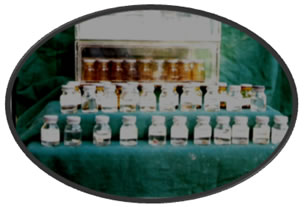
Histological stud: The lenses were divided into senile, diabetic and hypertensive groups and processed for histological study. As stated the lenses were preserved in 10% formalin.4,5,6 Then they were dehydrated in ascending grades of alcohol, cleared in toluene and embedded in paraffin wax. Then paraffin blocks were prepared from each lens5 and were given the same number as given to the patient. Later these blocks were cut, sections taken with rotary microtome, stained with Hematoxylin and Eosin and mounted on slides.7 These slides were finally numbered. These sections were observed under light microscope and the findings recorded.8 Photograph 1: Showing lens nuclei stored in plain bulbs in formalin
Estimation of total proteins About 100 of the lens nuclei were processed for estimation of total proteins after recording their colour. Proteins were calculated using Lowry’s method.9,10,11 The steps involved are as follows:- Homogenization: - homogenization was done using Potter Elvehjem Homogenizer9 in 2 ml Tris buffer (0.23 M, PH 7.85) containing 0.25x10⁻³ M EDTA and w/v adjusted to 5 gm%. Then the lens is centrifuged at 10,000 rpm for 1 hr and supernatant is used for estimation. 1) Lowry’s A – 2% Na2CO3 in 0.1 N NaOH 2) Lowry’s B1 – 0.5% CuSO4 : 5H2O 3) Lowry’s B2 – 1% Na Citrate 4) Lowry’s C – 1 ml Lowry’s B1 + 1 ml Lowry’s B2. It is freshly prepared and is diluted to 100 ml with Lowry’s A. Bovine serum albumin is used as a standard. Procedure: Three test tubes are taken and are used as Blank, Standard and Test. 0.1 ml of homogenate thus prepared is taken with the pipette in test tube. To the test tube first 1.9 ml of distilled water and then 2.5 ml of freshly prepared Lowry’s C reagent is added. Above solutions are mixed and kept for 15 minutes. Similar procedure is repeated for Standard and Blank test tubes. Then 0.5 ml of Folin Phenol Reagent is added to all three test tubes and kept for 45 minutes. Optical Densities of Blank, Standard and Test were noted using photoelectric colourimeter at wavelength 660 nm. Total proteins are calculated using the following formula:
Total protein O. D. of Test – O. D. of Blank conc. of Std/ml x vol of Std of = X homogenate O. D. of Std – O. D. of Blank volume of homogenate (mg/ml) The total lens proteins were expressed as mg protein/ gm wt of lens nucleus.
OBSERVATIONS Table 2: No. of cases in each colour group.
All 350 cases were sorted into four groups using Pirie’s classification. Male and female cases are separately tabulated. Table 3: Mean ages for each colour groups
Table no. III shows the mean age of cases in each colour group. The table shows increase in mean age for increase in colour of nucleus but this increase is statistically insignificant for both male and female and also between male and female. Table 4: Distribution of cases according to type of cataract
According to the history given by patients and study of their case papers, they were divided into five groups as shown. Almost 80% patients belong to senile group.
Table 5: Showing mean total protein concentration of each colour group of lens expressed as mg/gm wt of lens nucleus
Cataract is a protein condensation disease.12 The brown protein in cataractous lenses is restricted to nucleus of lens.3 Therefore we studied the levels of total protein in 100 cataractous lens nuclei and compared the levels between colour groups. Total protein concentration of lens nucleus shows a slight decrease as colour of nucleus advances. But this difference is statistically insignificant between each colour group and also not significant between male and female. In the present study, mean total protein concentration for human lens nucleus is found to be 432.86 + 23.38 mg/G wt of lens nucleus. DISCUSSION Colour of lens goes on increasing with age.5 Mean age for each colour group increases as colour deepens. However this increase in present study is not statistically significant. Colour of nucleus changes from pale yellow, dark yellow, yellow brown and dark brown to black.13,5 This occurs because of increase in water insoluble proteins of lens which accounts for its colour.3 Pale yellow lenses are seen in most younger patients while dark brown to black lenses are seen in elderly. Although age is the commonest risk factor for the darkening of lens colour, there are variable manifestations of intensities of colour in same age groups also which suggest a multifactorial etiology. Similar findings are recorded by Dorairaj et al8 and are compared in following table no VI.
Table 6: Comparison of age range (years) and colour of nucleus
Present study showed that no significant change in total protein concentration occurs in human cataractous lens nuclei. This finding is in total agreement with those by A. Spector et el14, Sanderson J et al15 and Pirie et al.3 In the bovine, rat and rabbit lenses there occurs a sharp increase in total protein concentration in nuclear region.14,16 In human lenses, however, there is progressive conversion of water soluble proteins into water insoluble protein and no change in total protein.11,114,16,17 and this process is more marked in nucleus which is most severely affected.3,9,13 The slight decrease in total proteins in brown lenses compared to yellow lenses may be because there is gradual loss of proteins, although small, from the more severely affected lenses. This occurs because of increasing hydration of lens and increasing calcium overload in cataractous lenses. The relatively low loss of total proteins indicates that the internal structures are relatively intact.15 We have measured all above parameters separately in male and female. Present study shows that no statistically significant differences exist in all above parameters between male and female. We have compared the values of total protein obtained by us with those of other workers. Table VII: Comparison of levels of mean total protein in lens nuclei
As shown in table VII our values of total protein are in agreement with those found by Bhat K.9 and Yadav S.2 Histologically, we observed the lens nuclei under light microscope after H and E staining. About 100 nuclei; 15 from each colour group which were the senile cataract lenses, 25 from hypertensive group, 10 from diabetic lenses and 5 from hypertensive and diabetic group were processed for the study. Sections of lens nuclei from patients belonging to colour group I showed fine well defined lamellations. The overall microscopic appearance was predominantly lamellar. Also in lenses belonging to group II and III we found well defined lamellations and at some places towards the adult nuclear region we found homogenous areas. In group IV lenses we found dense and homogenous areas. These sections also showed lot of serrations because of difficulty in cutting. The dense and homogenous areas were more marked towards the adult nuclear region although these observations were not consistently seen in all nuclei. These observations may be because of larger fibre cell size in embryonic and fetal nuclear regions compared to adult nuclear region. Also there occurs a process of fibre cell compaction which is more marked towards adult nucleus and inner cortex. These compacted fibre cells may become indistinguishable in these regions leading to its homogenous appearance.4 In lenses from diabetic individuals we found lamellated bands of lens fibres of differing densities. These bands may be associated with varied glucose level. 5 The variations in density may be because of alterations in cytoplasmic texture of adjacent fibre cells and may become a source for increased light scattering. Diabetes is considered to be one of the major risk factor for cataract17,18,19,21 and is known to accelerate the formation of cataract.1 The derangements of the various biochemical metabolisms occurring in lens in diabetes lead to early opacities that lead to scattering of light entering lens. In lenses from hypertensive individuals we found homogenous areas and certain areas of fine lamellations scattered with fine spaces. Similar histological observations are reported by Dorairaj et al.5 Systemic hypertension is again one of the major risk factors for cataract. Many studies have found systemic hypertension to be one of the important etiologies of cataract19,21,22,23 We searched for any sorts of opacities that may be responsible for light scattering in cataractous lens nuclei but at light microscopic level we could not locate any such opacities. In an attempt to search for this we also stained some of the sections with Toludine Blue. But the light microscopic appearance of the lens nuclei is similar in both H and E staining and Toludine Blue staining. Al-Ghoul K. J. et al have mentioned that only minor ultrastructural differences exist between the oldest fibre cells, located in nuclear region, in normal and cataractous lenses and that presence of extensive cellular damage and disruptions is not necessary for generation of nuclear opacities in aged lenses.24 We encountered numerous problems during sectioning of these lens nuclei. These difficulties were in the form of incomplete sections, folding of sections and numerous serrations in the sections. Sometimes the sections peeled off from the slides before mounting. The rotary microtome made a typical sound during cutting of these lens nuclei. These difficulties may be because of the hardening of the lens nucleus during aging. Costello et al (1992)25 in their study have stated that nuclear cataracts and nuclear cores of normal and human lenses are difficult to preserve as the tissue is dense, oval and not pliable. The high concentrations of cytoplasmic crystallins inhibit infusion of chemical fixatives and presents difficulties in sectioning. Gullapalli13 et al and Hu C et al26 have also found that hardness of the nucleus increases with deepening of nuclear colour. Heyworth et al27 have identified colour and age as major markers of nuclear hardness. Pau28 has found that brown or black colouration is related to maximum hardness but maximum hardening is not restricted to brown or black colouration. Although we were unable to measure nuclear hardness in this study; this is one of the physical parameters that show changes with age and colour4,16,26,29and needs to be studied.
REFERENCES
|
|
|||||||||||||||||||||||||||||||||||||||||||||||||||||||||||||||||||||||||||||||||||||||||||||||||||||||||||||||||||||||||||||||||||||||||||||||||||||||||||||||||||||||||||||||||||||||||||||||
 This work is licensed under a Creative Commons Attribution-NonCommercial 4.0 International License.