 Home
Home|
Table of Content Volume 12 Issue 3 - December 2019
Sexual dimorphism of maxillary sinus in Gujarat population CT - A retrospective study
Aakruti Rasiklal Parmar1*, Vipul V Solanki2
1Associate professor, Department of Anatomy, Government Medical College Bhavnagar 364001, Gujarat, INDIA. 2Assistant professor, Department of Anatomy, Government Medical College Bhavnagar 364001, Gujarat Email: aakrutiparmar@yahoo.com
Abstract Background: Variations in Morphological study of Maxillary sinus (MS) has anatomical, medico-legal and ethnic importance. Hence adult MS of both Sexes were studied radiologically (CT) Method: 20 Male and 20 female adults aged between 20-50 years were studied with CT images to measure the mediolateral, Supero–inferior and antero posterior dimensions and the volume of Maxillary sinus in both genders and obtained results were studied and compared statistically. Results: The mean value MLR in Males was 28.62 (S0±3.24) females 26.10 (±5.60), t test was 1.74 and P<0.01. The mean value of SIR was 39.11 (±4.66) in males, 34.20 (±5.43) in females t test 3.06 P<0.01. Mean value of APR in males 43.58 (±2.69) and females 37.02 (±3.03) t test – 7.24 P<0.01. Mean value of MLL in males was 28.02 (±4.03), females 24.11 (±5.17) t test 2.66 and P<0.01. Mean values SIL in males was 37.82 (3.32), females 34.12 (±5.60) t test. 2.54, P<0.01. Mean value of APL males was 41.78 (1.62) females 38.10(±1.98) t test was 6.43 and P<0.01. Mean values volume of right MS in Male 17.52 (±3.52) female 12.60 (± 4.14) t test 4.04, P<0.01. Mean value of left volume of MS in Males was 16.18 (±2.84), females 11.84 (± 3.88) t test was 4.03 P<0.01. Conclusion: This significant findings between Male and female MS has anatomical, anthropological and medico-legal importance in the adults. Key Word: MS Maxillary sinus CT = Computerised Tomography, VR=volume of right MS, VL=volume of left MS.
INTRODUCTION Maxillary sinus is present on both sides of the Norma frontalis, as they are made up of pneumatics bones hence they lightens the skull and contribute in the resonance of voice1. As they are pneumatic bones are delicate and more prone to break in cadaveric crania. Hence it is very difficult measure the any para-Nasal air sinus. Therefore computerised topographic study in living subjects was done to get exact measurements. The first PNS to develop is Maxillary sinus, (MS) is located in the right and left maxillary bones and consist of two pyramidal shaped air filled cavities lined by mucosa. The MS tend to appear at the end of the second embryonic month and complete by the age of 18 to 20 years of life.2,3 The shape and size of the MS varies amongst individuals, between genders and in various populations. The MS stabilise after the second decade of life and thus reliable measurements can be achieved by radiographic images (4). Owing to the nutritional status of foetus and mother, genetic factors of genders, environmental factors play vital role in the dimension of MS. Hence attempt was made to study the marphometry of MS in both sexes of adults, because marpho metric values of mesodermal derivatives are un-certain.
MATERIAL AND METHODS 20 male and 20 female adults aged between 20-50 years who were regularly visiting to government medical college hospital Bhavnagar Gujarat have been studied Inclusion criteria – The regular visitors did not have any pathology in PNS (maxillary sinus) and majority were healthy volunteers of both sexes were selected for study Exclusion criteria – The patients have pathology in PNS who undergone surgery of PNS children (below 18 years) and immuno compromised patients were excluded from the study
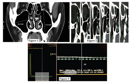
METHODS Non -contrast CT scan was performed to study the morphometry of the maxillary sinuses in both sexes using GE CT/e dual slice CT scanner (GE health care technologies Waukesha, WI, USA prior to the scan every patient was instructed to remove the metallic objects jewellery, hair pins etc, from the head to neck region and positioned on the CT Table in prone position. The patient neck was hyper-extended with the chin resting on the pad for stabilization. Pads were inserted on both sides of the head. The gantry was angulated to make it perpendicular to hard palate. 3mm thickness were used on preliminary scout view extending from anterior margin of frontal sinus to the posterior margin of sphenoid sinus with a reconstruction matrix size of 512X512 at 20 KV, 100Ma coronal CT was performed after instructing the patient to remain steady daring the entire procedure. The measurements like ML and SI were made maxillary sinus were in the widest position with the help of on screen linear management tool on the CT work station on the screen (Figure A). To measure the AP dimension of the maxillary sinus, the first and last appearance of the sinus was noted in the sequential coronal CT sections, and number of sections between them were selected. Finally selected sections were multiplied by 3 (thickness of a single section to find the AP of the sinus maxillary sinus volume (MSV) was Calculated by using the paint on slice tool on the work station. To define a volume, the outline of the sinus was traced manually on each slice of the image stack using the on scream mouse pointer in the coronal plane (Fig-B). Once tracing was compete, the work station automatically segmented the entire volume of the sinus from surrounding structure and the segmented portion could be visualized and manipulated in 3D. At this point, switching to the “histogram” view on the work station (fig-C) automatically reflected the volume of the sinus in cubic centimetres (CC) of both right and left maxillary sinus. The duration of study was from July 2006 to August 2008 (about two years). Statistical analysis - The obtained parameters were study in SPSS software computer.
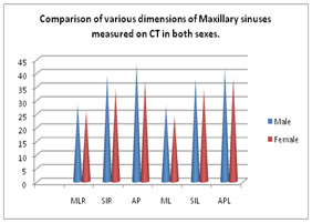
Figure A: Linear measurement of mediolateral and superoinferior dimensions of maxillary sinus; Figure B: “Paint on slice” tool; Figure C: Workstation showing the Maxillary sinus volume OBSERVATION AND RESULTS Table 1: Comparison of various dimension of maxillary sinuses measured on CT in both sexes
Table 2: Comparison of dimension of both sides in both sexes- mean value of VR mean value in male 17.52 (SD±3.52) females 12.60 (SD±4.14), t test value 4.04 and p value was highly significant (P<0.01). In VL – mean value in Males was 16.18 (SD±3.88) t test value was 4.03 and p value was highly significant (P<0.01).
Table 1: Comparison of various dimensions of Maxillary sinuses measured on CT in both sexes.
ML Right Medio lateral dimension of right side SIR Right Sup inferior dimension of Right side MX AP Right Antero posterior dimension of right sinus ML Left = Medio-lateral dimensions left maxillary sinus. SIL = Sup inferior dimension of left maxillary sinus, AP Left Anteriopost dimension of left maxillary sinus. Table 2: Comparison of dimensions Maxillary sinus of both sides in both sexes
VR – Volumes right maxillary sinus; VL – Volumes of left maxillary sinus
 DISCUSSION The present study of sexual dimorphism of MS, in Gujarat population. In the comparison of various dimensions of MS in both sexes– mean value of MLR in Males was 28.62 (±3.24) females 26.10 (±5.60) t test was 1.74 and P value was highly significant (P<0.01). In SIR mean values males was 39.11(±4.66) females 34.20 (±5.43) t test 3.06 P<0.01. In APR mean value of male 43.58 (±2.69) female 37.2 (±3.03) t test 7.24 (P<0.01) MLL mean value in males was 28.02 (±4.03) female 24.11 (±5.17) t test 2.66 P<0.01, SIL – mean value of males was 37.82 (±3.32), females 34.12 (±5.60) t test 2.54, P<0.01. In APL mean value of male was 41.78 (±1.62), female 38.10 (±1.98) t test 6.43 (P<0.01) (Table-1). In the comparison of volume of MS in both sides and both sexes VR – mean values males 17.52 (±3.52), females 12.60 (±4.14) t test -4.04, P<0.01. In VL mean value of Males was 16.18 (±2.84) in males 11.84 (±3.88) t test -4.03 and P value was highly significant (P<0.01). (Table-2) These findings were more or less in agreement with previous studies.5,6,7 This study will have great importance for gender determination because when any part of the crania or fragmented Skeleton is brought before anatomist or medico – legal expert it was very difficult to predict the gender because MS being delicate, fragile difficult remain intact. Hence only denser bones can remain intact by resisting fracture and incineration. The CT scan study of MS has of different dimensions is a robust method for sexual diamorphision9,10. The previous literature had mean values of MS measurements were 32, 25 and 35 mm in length, width and height respectively.11 It was established fact that overall size or dimensions of MS is larger in males than females.12 Moreover many factors are known to influence and modify the course of the bones development such as deprivation of raw materials, vitamins, hormonal imbalances and abnormal mechanical situations. But we have little idea how the genetic “blue print”. Contained in the nuclei of the osteoprogenitor cells is translated into action. These regional or ethnic variations might be due to ossification centres, because ossification centres may express their morphological individuality with astonishing persistence even though the bones which they represent have long ago ceased to be of any real functional importance. Hence variations have regional and morphological significance.
SUMMARY AND CONCLUSION The present study of sexual dimorphism of MS in Gujarat population is quite useful to anatomist, anthropologist and medico-legal expert but this demands further genetic, nutritional, environment, hormonal study because the factors which decide the Ossification is still un-clear.
REFERENCES
|
|
 This work is licensed under a Creative Commons Attribution-NonCommercial 4.0 International License.