 Home
Home|
Table of Content Volume 12 Issue 3 - December 2019
Guyons canal - Anatomical variations of ulnar nerve
Sharmada K L1, Kavya2*, Krishna Kishore3
1,3Tutor, 2Assistant Professor, Department of Anatomy, Bowring and Lady Curzon Medical College and Research Institute, Bangalore, Karnataka, INDIA. Email: kavs_084@yahoo.co.in
Abstract Background: Ulnar nerve is a very important nerve of the flexor compartment of the forearm and one of the vulnerable nerve due to its position. The Ulnar tunnel was first identified by Guyon hence the name. Ulnar nerve is known to give numerous unnamed muscular and articular branches; hence the study was undertaken to identify the detailed branching pattern of ulnar nerve in Guyons canal can help the operating surgeons to reduce complications leading to Ulnar nerve injury encountered during surgical procedures Materials and Methods: 18 human cadavers from Department of Anatomy, Bowring and Lady Curzon Medical College and Research Institute, Bangalore of unknown age and cause of death were included in the study. The skin, deep facia and the palmar aponeurosis were reflected and the ulnar nerve was identified and carefully dissected and branches identified. The ulnar nerve was identified by tracing its course from the medial epicondyle into the ulnar tunnel. Results and Conclusions: Symmetry in branching pattern was observed in 66.67% of the dissected specimen, in 5.55% of the dissected specimen premature bifurcation and trifurcation was observed. The understanding of the probable variation could help the surgeons avoid unwanted complications of nerve damage postoperatively. Key Words: anatomy, Guyon's canal, ulnar nerve, ulnar tunnel, variations
INTRODUCTION The ulnar nerve originates from the medial cord (C8-T1) and is a terminal branch of the brachial plexus. The ulnar nerve enters the palm by passing superficial to the flexor retinaculum lying lateral to the pisiform 12. Here, the ulnar nerve is covered by the volar carpal ligament 11. The area under this fascial band is termed as the ulnar tunnel. The Guyon's canal also called the ulnar tunnel is a fibro-osseous tunnel located on the antero-medial side of the wrist. 1 The roof of the canal comprises the superficial palmar carpal ligament and the palmaris brevis muscle, while the deeper flexor retinaculum and hypothenar muscles make up the floor. It is approximately 4 cm long, starting proximally at the transverse carpal ligament and terminating at the aponeurotic arch of the hypothenar muscles. The ulnar tunnel was first described by Guyon in 1861. McFarlane et al8 along with Enna et al9 proposed the name piso-hammate tunnel
AIM The Guyon's canal (ulnar tunnel) containing the ulnar nerve, artery and veins; is positioned in such a way that it is susceptible to neural injuries-mainly compression that requires surgery for the decompression of the ulnar nerve 2. This study was undertaken to study the variations of the ulnar nerve. Knowing the anatomy of the Guyon's canal is vital in diagnosing and treating ulnar tunnel syndrome, also known as Guyon's canal syndrome. The knowledge of the variations seen in the ulnar nerve in the Guyon's canal on cadaver specimens could be used as a reference during decompression wrist surgery.
MATERIALS AND METHOD 36 human cadavers from Department of Anatomy, Bowring and Lady Curzon Medical College and Research Institute, Bangalore of unknown age and cause of death were included in the study. Transverse incision 5cms above the wrist joint (incision 1), A vertical incision was made from the middle of incision 1 to the base of the middle finger (incision 2), A transverse incision from the base of the index finger was made, up to the base of the little finger (incision 3) and A vertical midline incision extending from the midline of the line join the medial and lateral epicondyle to the midpoint of incision 1 (incision 4). The skin, deep facia and the palmar aponeurosis were reflected and the ulnar nerve was identified and carefully dissected and branches identified. The ulnar nerve was identified by tracing its course from the medial epicondyle into the ulnar tunnel.
RESULTS
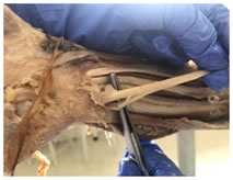
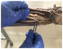
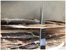
Figure 1 Figure 2 Figure 3 Figure 1: Branching of Ulnar Nerve in Guyons canal into Superficial and Deep branch; Figure 2: Premature bifurcation of the Ulnar Nerve; Figure 3: Trifurcation of Ulnar Nerve
Symmetry in branching patterns was seen in 94.4% of the specimens studied. The prevalence of premature bifurcation of the ulnar nerve was thus calculated to be 2.77% The prevalence of premature trifurcation of the ulnar nerve was thus calculated to be 2.77%
DISCUSSION The ulnar nerve passes through the ulnar tunnel as it runs towards the hand to provide motor and cutaneous innervation to the digits where it is prone to entrapment syndromes. The goal of this study was to gather cadaveric data on the prevalence of variations in the ulnar nerve as it divides into its branches at the ulnar tunnel, difference wasn’t seen in the symmetry of branching pattern of both sides when compared. About 94.4% of the specimens showed symmetry in branching, of the ulnar nerve in the Guyon’s canal. In 2.77% of the specimens showed variation in the position of bifurcation of the ulnar nerve, the nerve split into its superficial and deep branches prior to the ulnar tunnel. In 2.77% of the specimen trifurcation of the ulnar nerve was seen into two cutaneous and one muscular branch. The clinical significance of this study includes the variations in the ulnar nerve and the role it plays in ulnar nerve entrapment. Bozkurt et al. stated that the most common site for compression is the distal end of the tunnel along with suggested causes for the nerve compression.4 Murata et al. observed that in most cases nerve compressions are idiopathic with trauma finishing second.5 A rare cause of compression is anatomic variations of muscles in the Guyon’s canal.6 As the symmetry is high, the risk of injury in the area is not increased on either side and this is significant when seen from a surgical point of view.
CONCLUSION The ulnar nerve shows a variety of differences in the branching pattern. The common variations seen include premature bifurcation of the ulnar nerve into its cutaneous and deep branches and the trifurcation of the nerve into the deep and superficial divisions. A thorough understanding of the probable variations can help surgeons reduce complications during decompression surgeries.
REFERENCES
|
|
 This work is licensed under a Creative Commons Attribution-NonCommercial 4.0 International License.