 Home
Home
|
Table of Content Volume 13 Issue 1 - January 2020
Study of metopic suture in adult human skulls of Marathwada region
Vaishali Sitaram Kirwale¹, Shivaji B Sukre²*
1Assistant Professor, 2Professor & HOD, Department of Anatomy, Government Medical College, Aurangabad, Maharashtra, INDIA. Email: drsukresb@yahoo.co.in
Abstract Background: Metopic sutures are vertical sutures between two halves of the frontal bone, it extends from anterior fontanelle (bregma) to the nasion. When the complete suture persists between bregma and nasion it is called as metopism whearas if only small part persists then called as incomplete metopic suture. Materials and Methods: The present study was carried out by using 116 adult dry human skulls. The samples were collected from the Department of Anatomy, Government Medical College Aurangabad. The skulls were carefully examined for the presence of metopic suture and it’s morphological variations Results: In the present study 116 adult dry human skulls we’re studied out of which 27 (23.27%) skulls showed the persistent metopic suture. The complete metopic suture (metopism) was noted in 4 (3.44%) of cases. 24 (20.68%) of skulls revealed persistent incomplete metopic sutures. Among the morphological variations Linear type was observed in 9 (7.75%) of cases. 7 (6.03%) skulls showed the presence of U shaped variation. V shaped variation was seen in 4 (3.44%) skulls. The Y shaped 2 (1.72%) and Bilinear varient were also noted in 2 (1.72%) skull. The other variations like H shaped, inverted U shaped did not found in the present study. Conclusion: The persistent metopic sutures commonly misdiagnosed as fracture so that radiologists, forensic expert and neurosurgeon should have knowledge of metopic suture and it’s variations. Key Words: metopic suture, metopism.
INTRODUCTION Metopic suture connects the two frontal bone in developmental stage. It is a kind of dentate suture and is seen in infants. It usually closes at an age of 6 years when the two frontal bones fuses together. Sometimes the metopic suture persists in adults even after fusion and it is called as persistent metopic suture. The persistent metopic suture may occur as two forms. The suture from bregma (anterior frontanellae) till the nasion called as complete metopic suture or metopism. Incomplete(Partial) suture extending from nasion not till bregma or from bregma not till nasion. Metopic suture normally closes by fifth or sixth year¹. Hamilton² stated that the metopic suture disappears by seventh year of life. Fusion of this suture commences at the anterior fontanelle i.e., bregma and terminates at nasion. Williams³ claims that the frontal bones are separated by the metopic suture at birth; this is obliterated by 6-8 year. Keith⁴ stated that the metopic suture disappears at the end of the first year, or in the beginning of the second year of life. A.K. Dutta⁴ stated that at birth the 2 halves of the frontal bone are separate as the metopic suture, replaced by bone at the age of 2 years. Remnants of the metopic suture may persist in some skulls at glabella. So the upper limit for the persistence of metopic suture may extends upto 8 years. The metopic suture may be mistaken as fracture of skull at frontal bone by an inexperienced forensic expert. This anatomical variation should be known to neurosurgeon while performing frontal craniotomy. The present study is undertaken to find out the incidence of metopic suture among Marathawada region. The knowledge of metopic suture is important in order to not to mistaken it as fracture also neurosurgeons, radiologists should have knowledge about metopic suture and it’s variations.
MATERIALS AND METHODS The present study was carried out by using 116 adult dry human skulls. The samples were collected from the Department of Anatomy, Government medical college Aurangabad during the year 2019.The skulls were carefully observed for the presence of metopic suture by naked eyes. Complete and incomplete metopic sutures were recorded along with the morphological variation of their shapes. The length of the complete metopic suture were also recorded by using thread spread from bregma to nasion. The above obtained data was properly recorded and analysed. The findings if present study were compared with the previous studies held by various authors.
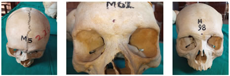
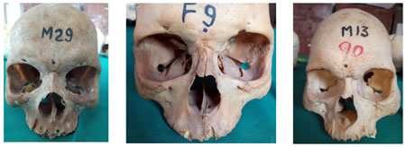
RESULT In the present study in total 116 adult dry human skull were studied, out of which 23.27% of skulls shows the presence of metopic suture whereas the rest 85.34% of skull are of without metopic suture. Among total 23.27% of skulls with metopic suture 3.44% of skull shows presence of complete metopic suture i.e metopism (Photo-1), while 20.68% of skulls shows presence of incomplete metopic suture. Five types of variation in the shape of metopic suture were noticed in the present study. The Linear incomplete metopic suture (Photo-2)were observed in 9 (7.75%) of skulls. The U shaped incomplete metopic suture (Photo-3) were noted in 7 (6.03%) of skulls. The V shaped suture (Photo-4) were found I 4 (3.44%) of skulls. The Y shaped suture (Photo-5)were noticed in 2 (1.72%) and Bilinear suture (Photo-6) in 2 (1.72%) of skulls.
TABLE 1: Metopic suture findings in present study
Photo 1 Photo 2 Photo 3
Photo 4 Photo 5 Photo 6 Photo 1: Complete Metopic suture (metopism); Photo 2: Linear variety of incomplete metopic suture; Photo 3: U shaped incomplete metopic suture; Photo 4: V shaped incomplete metopic suture; Photo 5: Y shaped incomplete metopic suture; Photo 6: Bilinear incomplete metopic suture. DISCUSSION The incidence of metopic suture compared with the findings of previous workers. (Table No.2) Overall incidence varies from 1 to 10%. The lowest incidence was reported by Breathnach6 which was 1% in Africans and also in Australian population (1%) studied by Bryce7. The highest incidence was observed by the Woo8 which was 10% among Mongolian population, also the European population studied by Breathnach6 and Scttish population studied by Bryce7 shows higher rates of metopism which was 7-10% and 9.5% respectively. In the present study incidence of metopism is 3.44% among the Marathwada region in Maharashtra state. The findings of present study resembles with the Das et al9which was 3.31% and also with the Ajmani et al10 which was 3.4%. This study describes the incidence incomplete metopic suture of about 20.68%. The Das et al9 reported the incidence of incomplete metopic suture of about 17.57%, by Agrawal et al11was 35.5%, by Ajmani et al10 was 31.57% and by Shanta Chandrasekaran12 was found to be 40%. Thus the findings of present study in relation to the incidence of incomplete metopic suture correlates with the findings of Das et al9 The linear incomplete metopic suture were noted in 7.75% of cases in present study whearas Agrawal it al11 reported it in 23.12% of skulls, 17% by Shanta Chandrasekaran12. In this study the incidence of U shaped suture is 6.03% whearas Shanta Chandrasekaran12 observed it to be 15%. The incidence of V shaped suture is 3.44% in this study. The Das et al9 observed V shaped suture in 1.01%, Agarwal et al11in about 3.25%, Ajmani et al10 in 0.49% and S.chandrasekaran12 by 7.5%. The incidence of V shaped suture of present study (3.44%) resembles with the findings of Agarwal et al11 (3.25%). The incidence of Y shaped suture is 1.72% in our study which is similar to the findings of Inderjit and shah13 and Agarwal et al11 which was 1.25% and 1.96% respectively. The incidence of Bilinear suture in the present study is 1.72% .The other variations like H shaped, inverted U shaped did not noticed in the present study. The mean suture length of complete metopic suture was reported to be 121.4mm and 123.1mm by the skrzat et al whearas it is 120.75mm in the present study. TABLE 2: Incidence of metopism by various authors
CONCLUSION The present study was conducted for the incidence of metopic suture in 116 adult dry human skulls. The suture (metopism) was detected in 3.44% of skulls and incomplete metopic suture was present in 20.68% of skulls. This anatomical knowledge and morphological variation of metopic suture is usefull for the surgeons, forensic experts and radiologists as it can be commonly mistaken as fracture of frontal bone. The neurosurgeons should also aware of these persistent suture while performing frontal craniotomy.
REFERENCES
Policy for Articles with Open Access: Authors who publish with MedPulse International Journal of Anatomy (Print ISSN: 2550-7621) (Online ISSN: 2636-4557) agree to the following terms: Authors retain copyright and grant the journal right of first publication with the work simultaneously licensed under a Creative Commons Attribution License that allows others to share the work with an acknowledgement of the work's authorship and initial publication in this journal. Authors are permitted and encouraged to post links to their work online (e.g., in institutional repositories or on their website) prior to and during the submission process, as it can lead to productive exchanges, as well as earlier and greater citation of published work.
|
|
 This work is licensed under a Creative Commons Attribution-NonCommercial 4.0 International License.