 Home
Home
|
Table of Content Volume 13 Issue 1 - January 2020
Neeraj Master1, Nisha Parmar2, Deepa S Gupta3*
1,2 Tutor,3 Professor & Head, Department of Anatomy, Surat Municipal Institute of Medical Education and Research (SMIMER), Surat-395010, Gujarat, INDIA.
Abstract Background: Biceps brachii muscle has two heads of origin: short head and long head. Bicep brachii is reported to have supernumerary head or 3rd head of origin. Such supernumerary heads of origin and associated variations in neural innervations of arm muscles are important for clinicians. Aim: To assess the incidence of supernumerary head of biceps brachii muscle and associated variation in the nerve supply in the cadavers of south Gujarat. Materials and Method: Study was done on 96 upper limbs belonging to 48 embalmed cadavers (M: F = 27: 21) donated in the department of anatomy. Proper labelling of the upper limbs for number, male/female, right/left side was done. Dissections of the all upper limbs were done as per standard dissection guidelines. Biceps brachii muscle was examined for origin, insertion, nerve supply and variation like extra head of biceps if any. Nerve supply of the all the muscles of the front of the arm were accessed. The neuromuscular variations if any were noted and photographed. Results: Out of total 96 upper limbs, supernumerary or extra head of bicep brachii was observed in eight (8.33%) cases (3 female: 5 male cases). Five cases were of left side and three were of right side. Out of 8 upper limbs, 6 upper limbs were associated with variations in the nerve supply of the front arm muscles. In seven cases, 3rd head was arising from the middle of the shaft humerus below the coracobrachialis insertion. In one case, both the heads were having 3 different slips of origin. In 3 cases extra head was supplied by median nerve and in 5 cases it was supplied by musculocutaneous nerve. Musculocutaneous nerve was absent in two of above cases. Results were compared with previous studies and discussed. Conclusion: Awareness regarding the incidence of muscular variations like extra head of biceps brachii and innervations of arm will help the surgeons, anaesthetists and neurophysicians working in this region. Key Words: Supernumerary head of biceps, 3rd head of bicep brachii, musculocutaneous nerve, median nerve, communications, neuromuscular variations
INTRODUCTION Bicep brachii is the muscle of the front of the arm having two heads of origin. Short head of biceps brachii originates from the tip of the coracoids process in association with coracobrachialis muscle and long head of biceps arises from the supraglenoid tubercle of the scapula. Both head unite to form the belly and inserted on the radial tuberosity. Few fibres form the apponeurosis and merge with deep fascia of the front of the forearm medially. Biceps brachii muscle is the main supinator of forearm in flexed elbow as well as flexor of elbow joint 1. Studies done by different researcher have shown that bicep brachii shows frequent anatomic variations in the form of supernumerary head mostly arising from the humerus near coracobrachialis insertion and brachialis origin, and/or from medial intermuscular septum. In few cases it also arises from the tendons of pectoralis major or deltoid, or from articular capsule or from greater tubercle or coracoids process. Such head then join the common belly or to the belly of long head or short head or to the apponeurosis of biceps brachii 2,3,4,5. Muscles of the front of the arm including biceps brachii are supplied by the musculocutaneous nerve 1. Many studies have shown that communications between musculocutaneous nerve and median nerve as well as variations in innervations of front of arm muscles are common 6, 7, 8. Muscles of the front of the arm along with the variations like supernumerary/extra head (3rd head) of bicep brachii are supplied by musculocutaneous nerve 3 or sometimes branches from communications between musculocutaneous nerve and median nerve or directly from median nerve 7. So, the present study was conducted to assess the incidences of supernumerary or 3rd head of biceps brachii and associated variations in the innervations of the front of arm muscles in the cadavers of South Gujarat region.
AIMS
MATERIALS AND METHODS Study was done in the Anatomy Department, Surat Municipal Institute of Medical Education And Research (SMIMER), Gujarat, India, on 96 upper limbs belonging to 48 embalmed cadavers (M: F = 27: 21) donated over previous four years. Labelling of the upper limbs were done based on number, side (right or left) and sex of the body (male or female). For example case 5MR denotes: five number case, male body & right upper limb. Dissections of the all upper limbs were done as per dissection guidelines given by Cunningham’s manual of Practical Anatomy 9. Biceps brachii muscle was exposed. Origin and insertion of the muscle, its nerve supply, variation like extra head of biceps if any were carefully dissected and noted. Nerve supply of the all the muscles of the front of the arm were accessed. The morphological variations regarding musculocutaneous nerve and median nerve in arm if any were noted and photographed.
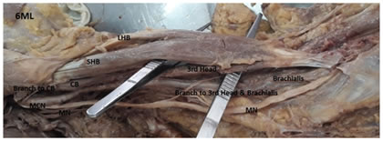
RESULTS Out of 96 upper limbs (48 cadavers) dissected, supernumerary or extra head of bicep brachii was observed in eight cases (6ML, 11FL, 18MR, 24FL, 28FR, 33ML, 35MR, 42ML). Three cases were of female and five cases were of male cadavers. Five cases were of left side and three were of right side. All the cases were unilateral. Amongst 8 upper limbs which were found to have supernumerary or 3rd head of biceps brachii variation, 6 upper limbs (6ML, 11FL, 18MR, 24FL, 33ML, 42ML) were associated with variations in the nerve supply of the front arm muscles and 2 upper limbs (28FR, 35MR) were have normal nerve supply. Total 21 upper limbs out of 96 upper limbs dissected had showed some form of variations in nerve supply of the front of the arm muscles. We did not find any variations in the insertion of the biceps brachii. Details of variations of all eight limbs (6ML, 11FL, 18MR, 24FL, 28FR, 33ML, 35MR, 42ML) were as follows: CASE 1: 6ML: Extra 3rd head was arising from upper shaft of humerus, was thick and united with rest of the muscle to form single tendon for insertion. MCN was not piercing the coracobrachialis and was giving a branch to it which was also supplying to biceps brachii. After it MCN fused with MN. 3rd head of biceps and brachialis was supplied by braches from MN. (Figure 1) Figure 1: 6ML: 3rd head arising from the upper shaft humerus, MCN fuses with MN after supplying CB and BB. 3rd head and brachialis were supplied by MN branch. (MCN= musculocutaneous nerve, MN= median nerve, SHB=short head of biceps, LHB= long head of biceps, CB= coracobrachialis, BB= biceps brachii)
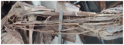
CASE 2:11FL: Extra 3rd head of biceps was arising from the middle of the shaft. MCN was not piercing the corcobrachialis and supplied by small branch. MCN later fused with MN in the middle of arm after supplying all the muscles of arm. MN did not supply any forearm muscle. (Figure 2) Figure 2: 11FL: 3rd head arising from middle of the shaft humerus, musculocutaneous nerve fused with median nerve after supplying arm muscles including 3rd head of biceps. (MCN= musculocutaneous nerve, MN= median nerve, SHB=short head of biceps, LHB= long head of biceps, CB= coracobrachialis, BB= biceps brachii)
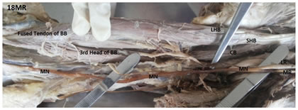
CASE 3:18MR: Extra 3rd head of biceps was arising from the lower anteromedial shaft below the insertion of coracobrachialis and above the origin of brachialis. MCN was absent and lateral chord was supplying coracobrachialis and then fused with MN. Rest of the muscle of arm including third head of biceps was supplied by branches arising from MN which was formed by three roots. The lateral cutaneous nerve of forearm was from median nerve. (Figure 3)
Figure 3: 18MR: shows 3rd head arising from the middle of the shaft humerus, absent musculocutaneous nerve and median nerve supplies all the muscles of the arm. (MCN= musculocutaneous nerve, MN= median nerve, SHB=short head of biceps, LHB= long head of biceps, CB= coracobrachialis, BB= biceps brachii)
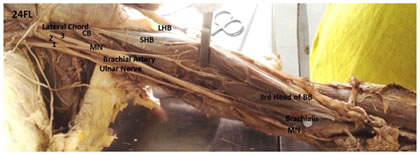
CASE 4: 24FL: Extra 3rd head of biceps was arising from the lower shaft of humerus below the insertion of coracobrachialis. Musculocutaneous nerve (MCN) was absent and all the flexor muscles of the arm including 3rd head were supplied by branches from median nerve (MN) which was having three roots of origin. The lateral cutaneous nerve of forearm was from median nerve. (Figure 4)
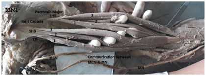
Figure 4: 24FL: shows 3rd head of biceps brachii(BB) arising from the shaft humerus, absent musculocutaneous nerve and median nerve formed by three roots(1,2,3) and supplies all the muscles of the arm. (MCN= musculocutaneous nerve, MN= median nerve, SHB=short head of biceps, LHB= long head of biceps, CB= coracobrachialis, BB= biceps brachii) Case 5: 33ML: It showed that the short head of biceps had 3 slips and the long head of biceps also had 3 slips which united to form a single tendon of insertion. Origins of both head were from normal anatomical sites as a single tendon of origin. Normal pattern of two fusiform bellies seen in biceps brachii muscle was absent. All of them were supplied by muculo-cutaneous nerve and it was also giving communication branch to median nerve in the middle of the arm. (Figure 5) Figure 5: 33ML showing both the short head and long head of biceps had three separate slips giving the muscle six head like appearance. (MCN= musculocutaneous nerve, MN= median nerve, SHB=short head of biceps, LHB= long head of biceps, CB= coracobrachialis, BB= biceps brachii)
CASE 6: 42ML: Extra 3rd head of biceps brachii was arising from the middle of the shaft of humerus below the insertion of coracobrachialis and then fused with common tendon of insertion. Musculocutaneous nerve was not piercing the coracobrachialis. MCN was supplying all the muscles of the front of arm including extra head of biceps brachii. MN did not supply any of the arm muscle. (Figure 6)
Figure 6: 42ML: 3rd head arising from the middle of the humerus. MCN was supplying all the muscles. (MCN= musculocutaneous nerve, MN= median nerve, SHB=short head of biceps, LHB= long head of biceps, CB= coracobrachialis, BB= biceps brachii) Remaining 2 upper limbs (28FR, 35MR) also showed extra 3rd head of origin from the lower shaft humerus and were having normal anatomical nerve supply and blood supply. Table 1: Incidence of supernumerary/extra 3rd head of biceps brachii in different studies
( - ) = values not specified/ data not mentioned
DISCUSSION Present study showed presence of supernumerary head/3rd head of biceps brachii in 8.33% cases (8 limbs out of 96 upper limbs) of south Gujarat population. Standard textbook showed 10% variation in bicep brachii 10. Various studies have shown differences in the incidences of supernumerary head of biceps brachii based on region and races ranging from 2.0 % to 37.5%. (Table1). It was found to be, 8% in Chinese 12,10% in Europeans 12, 13-21% in Japanese 2, 8-21% in South Africans 3,11,15% in Turkish 4, 37.5% in Colombians 5, and 2%-10% in Indians 13,14,15,16. Higher results showed in few studies done were as a result of calculation of the cadaver number and not the numbers of upper limbs examined or due to small sample size. Results of our study were in mid-range of other sub continental studies but were in higher range when compared to Indian studies.We did not found variations like absence of the muscle or absence of any head of origin or variations of insertion in any case. Out of total 48 cadavers, 27 were male and 21 were female cadavers. No significant male to female (5 male: 3 female) differences or left to right limb (5 left limb: 3 right limb) difference were found in this study. All cases were of unilateral limbs. Such variations were mention by Asvat et al3 (in 6 male, 1 female and 9 bilateral male, total 25 limbs), Kosugi et al2 (in 23 male,18 female, 13 bilateral male and 4 bilateral female, total 75 limbs ) and Rincon et al5 (in 3 male, 1 female and 2 bilateral male, total 8 limbs). Many studies have shown that biceps brachii may have supernumerary head of origin in the form of 3rd head, 4th head or even more 2, 11, 17. Nasr AY et al had found the origin of the fourth head of biceps brachii from the articular capsule of shoulder joint in 1 limb and from the coracoid process of scapula in the another 1 limb. These extra-head then fused either with the long head and/or united with the short head 18. In one case (33ML) of this study, we found that the short head of biceps brachii after originating from the coracoids process as a single tendon was separated as 3 different slips or bellies. Similarly the long head of biceps after originating as a single tendon, separated into three separate slips/bellies. These bellies then unite with the corresponding heads or common tendon before the insertion. Presence of such 6 different numbers of slips of biceps brachii was appearing as six headed muscle and was unique of this study except for double short head of biceps arising from coraciod process or double long head arising from the shoulder joint capsule reported in some studies11, 16. Greig HW et al11 had founded 28 cases with extra heads of origin. They had described: (A): 18 cases of accessory humeral head as most common arising from the middle of the shaft of humerus below the coracobrachialis insertion and brachialis origin. Out of which 11 cases had three heads (tricipital), 5 cases had 4th head (quadricipital) and 2 cases had five heads of origin (pentacipital) arising from humerus. (B) In 7 cases they found accessory head of muscle origin or double head and (C) in 3 cases the head of origin were replaced and arisen from bicipital groove or capsule of shoulder joint. In present study 7 cases were having 3rd head arising from the middle of the shaft humerus similar to type A of Greig et al. In one case there were 3 slips for short head and 3 slips for long head appearing as unique hexacipital origin. Out of eight cases of supernumerary heads, in five cases 3rd head along with all the front arm muscles were supplied by musculocutaneous nerve and in three cases median nerve was supplying the 3rd head. Musculocutaneous nerve was completely absent in two of the above cases. Kosugi K et al2 had mention that 43 out of the 75 limbs having extra head of biceps brachii (57.3%) were having communication between the musculocutaneus nerve and the median nerve. In present study we found 8 cases of extra head of origin of biceps brachii and 6 out of them (75%) were associated with variation in nerve supply, which was high in accordance with results of Kosugi et al. Variations in MCN and MN in upper arm were documented by various studies 6,7,8. We had also found 21 cases (21.88%) of such variations out of 96 upper limbs. Le minor8 had classified such variations in five types. Type1: no communications between MCN and MN, Type 2: mid arm communication between MCN and MN, Type 3: lateral root joins MN via MCN in the arm, Type 4: MCN arise from MN in arm and Type 5: absent MCN and arm muscles supplied by branches from MN. Such type of combined neuromuscular variations had been mention by Kosugi et al2 in 24(32%), 12(16%), 5(6.7%) and 2(2.66%) cases out of 43 cases, similar to variations mention in le minor classification. In present study, out of total 8 positive cases of the supernumerary or extra 3rd head of biceps brachii, three cases (28FR, 35MR, 42ML) were of type 1, two cases (11FL & 33ML) were of type 2, one case (6ML) was of type 3 and remaining two cases (18MR and 24FL) were of type 5 of Le minor classification. In later two cases (18MR, 24FL) musculocutaneous nerve was absent and all the arm muscles including 3rd head of biceps brachii were supplied by median nerve. Absent MCN with extra 3rd head of biceps brachii found in two cases of this study were rare occurrence and very few researchers have documented it19,20,21. Anatomical variations seen in present study can be explained by the process of development. As the upper limb bud is form mesenchyme migrates into it to form ventral and dorsal muscle masses. Ventral muscle mass give rise to muscles of front of arm including biceps and it is innervated by ventral primary rami. Combinations between ventral segmental braches give rise to median nerve and musculocutaneous nerve formation. Peripheral nerve axons are guided to their target organ by apical growth cone and correct path-finding is influenced by various tropical substances. Alteration in signalling between mesenchymal cells and neuronal growth cone during 4th to 7th week of development results in variations in nerve supply as well as formation of supernumerary heads 22. Testut23 had described such variations of 3rd head of biceps brachii as distal translocated part of insertion of brachialis muscle from the ulna to radius. Phylogenically the 3rd head of biceps brachii arising near the insertion of coracobrachialis muscle found in present study was described as remnant of the long head of the coracobrachialis-as an ancestral hominoid condition by Sonntag 24.It is likely that such supernumerary heads are asymptomatic in most of the cases and present as an incidental findings either found during anatomical dissection or encountered during the radiological imaging and/or during surgery of the arm. Kosugi K et al2 had stated that the presence of a supernumerary head seemed to affect the course and branching of the musculocutaneus nerve. The presence of such supernumerary head of the muscle may cause compression of the neurovascular structure and may lead to variation of normal mechanical action 16, 19, 23.Knowledge of variations regarding extra supernumerary head of biceps brachii associated with variations in the innervations of front of the arm muscles is interesting not only to anatomists but also to the clinician, in other words, not only by the phylogenetic viewpoint but also by the surgical viewpoint, particularly when it is related to the course of the musculocutaneus nerve2. So, the surgeons, anaesthetists and neurophysicians who are dealing with the trauma surgeries, nerve block or nerve compression management in upper arm should keep this in mind. Present study will aid to the better understanding in the relationship of the communications between MCN and MN and the existence of the supernumerary head of biceps brachii.
CONCLUSION In present study, we found that biceps brachii infrequently showed extra head of origin mostly in the form of 3rd head arising from the middle of the shaft of humerus and was associated with variations in the innervations of the arm muscles. Awareness regarding the incidence of such neuromuscular variations will help the surgeons, anaesthetists and neurophysicians working in this region.
REFERENCES
Policy for Articles with Open Access: Authors who publish with MedPulse International Journal of Anatomy (Print ISSN: 2550-7621) (Online ISSN: 2636-4557) agree to the following terms: Authors retain copyright and grant the journal right of first publication with the work simultaneously licensed under a Creative Commons Attribution License that allows others to share the work with an acknowledgement of the work's authorship and initial publication in this journal. Authors are permitted and encouraged to post links to their work online (e.g., in institutional repositories or on their website) prior to and during the submission process, as it can lead to productive exchanges, as well as earlier and greater citation of published work.
|
|
|||||||||||||||||||||||||||||||||||||||||||||||||||||||||||||||||||||||||||

 This work is licensed under a Creative Commons Attribution-NonCommercial 4.0 International License.