 Home
Home
|
Table of Content Volume 13 Issue 2 - February 2020
Abdominal aorta - Bilateral arterial variations
K Satheesh Naik1*, M Gurushanthaiah2
1Assistant professor, Department of Anatomy, Viswabharathi Medical College and General Hospital, Penchikalapadu, Kurnool, Andhrapradesh, INDIA. 2Professor, Department of Anatomy, Basaves hwara Medical College and Hospital, Chitradurga, Karnataka, INDIA Email: nsatheesh43@gmail.com
Abstract Background: The abdominal aorta is an important artery in various abdominal surgeries. Hence, the aim of this study was to observe the variations in the branching pattern of abdominal aorta in cadavers. Material and Methods: We Dissected 40 cadavers of both the sex for Medical under graduates and came across the variations in branching pattern of abdominal aorta in about 3 male cadavers, bilaterally and variations were photographed. Results: In Laparoscopic surgeries and kidney transplantation Variations in the branching pattern of the aorta was clinically important. We observed bilateral accessory renal arteries arising from abdominal aorta; coeliac trunk gives rise to a common arterial trunk, which divides into left inferior phrenic and Left middle suprarenal arteries. Left superior suprarenal artery was arising from left inferior phrenic artery and left inferior suprarenal artery normally arising from left renal artery. We also studied the right inferior phrenic artery arising from abdominal aorta below the origin of coeliac trunk, and gives rise to right superior suprarenal artery. Right inferior suprarenal artery was arising from right accessory renal artery; right middle suprarenal artery was absent. We also observed Right gonadal artery was arising from ventral surface of abdominal aorta and left gonadal artery was arising from right accessory renal artery. Conclusion: The knowledge of arterial variations in radio diagnostic interventions and legating blood vessels in abdominal surgeries is useful for the surgeons. Key Wards: Abdominal aorta, coeliac trunk, Inferior phrenic, Accessory renal and gonadal arteries.
INTRODUCTION At the level of lower border of 12th thoracic vertebra thoracic aorta continues as abdominal aorta after passing through the median Osseo aponeurotic hiatus in the diaphragm. It continuous downwards up to the level of the fourth lumbar vertebra then bifurcates into the right and left common iliac arteries. The branches of abdominal aorta divided into anterior, lateral and dorsal branches 1. The celiac trunk is the first anterior branch of abdominal aorta arises at the level of T12 – L1 below the aortic hiatus and gives rise to the left gastric, Splenic and common hepatic arteries by supplying forgut derivatives. Superior mesenteric artery arises 1cm below the coeliac trunk, at the level of the L1-L2 intervertebral disc and supplies midgut derivatives. At the level of L3 vertebra the inferior mesenteric artery arises from the anterior aspect of the abdominal aorta and supplies hindgut derivatives.2 The lateral branches of the aorta, i.e. renal arteries and gonadal vessels supply the urogenital system. The posterolateral branches, i.e. inferior phrenic arteries and the lumbar arteries supply the body wall, inferior aspect of the diaphragm and posterior abdominal wall 3. Variations in abdominal aorta and its branches are frequently observed and they occur due to embryological developmental changes.
MATERIALS AND METHODS 40 cadavers of both the sex were dissected for medical under graduates in Basaveshwara medical college, Chitradurga, Karnataka. We observed the following bilateral variations in the branching pattern of abdominal aorta in 3 male cadavers; the data obtained was compared with the previous studies.
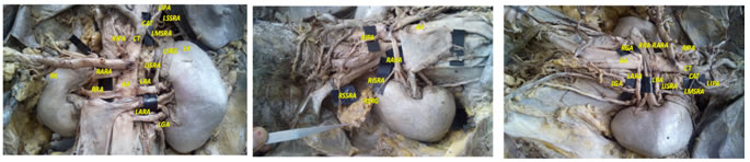
RESULTS We observed left accessory renal artery was arising from abdominal aorta below the origin of left renal artery; right accessory renal artery was arising from abdominal aorta above the origin of right renal artery. The Coeliac trunk was arising from abdominal aorta and gives rise to a common arterial trunk, which was dividing into left inferior phrenic artery and ascending up, to supply diaphragm and left middle supra renal artery coursing obliquely down, to supply left supra renal gland. The left superior suprarenal artery was arising from left inferior phrenic artery and left inferior suprarenal artery was normally arising from left renal artery (Figure.1). We also observed the right inferior phrenic artery was arising from abdominal aorta below the origin of Coeliac trunk and coursing obliquely up, to supply diaphragm and gives rise to superior supra renal artery, right inferior supra renal artery was arising from right accessory renal artery and coursing upwards to supply suprarenal gland, right middle supra renal artery was absent (Figure.2) Right gonadal artery was arising from ventral surface of abdominal aorta and left gonadal artery was arising right accessory renal artery and going to supply both the testis (Figure.3). Distribution of variations in the branching pattern of abdominal aorta shown in the table: 1
Figure 1 Figure 2 Figure 3 Figure 1: Showing origin of AA: Abdominal aorta, CT: Coeliac trunk, CAT: Common arterial trunk, LIPA: Left inferior phrenic artery, LSSRA: Left superior suprarenal artery, LMSRA: Left middle suprarenal atery, LISRA: Left inferior suprarenal artery, LRA: Left renal artery, LARA: Left accessory renal artery, LK: Left Kidney, LSRG: Left suprarenal gland, LGA: Left gonadal artery, RIPA: Right inferior phrenic artery, RARA: Right accessory renal artery, RRA: Right renal artery and RK: Right Kidney; Figure 2: Showing origin of RIPA: Right inferior phrenic artery, RSSRA: Right superior suprarenal artery, RARA: Right accessory renal artery, RISRA: Right inferior suprarenal artery and also AA: Abdominal aorta and RSRG: Right supra renal gland; Figure 3: Showing origin of AA: Abdominal aorta, CT: Coeliac trunk, CAT: Common arterial trunk, LIPA: Left inferior phrenic artery, LMSRA: Left middle suprarenal atery, LISRA: Left inferior suprarenal artery, LRA: Left renal artery, LARA: Left accessory renal artery, LGA: Left gonadal artery, RIPA: Right inferior phrenic artery, RARA: Right accessory renal artery, RRA: Right renal artery and RGA: Right gonadal artery.
Table 1: Showing the Variations in the branching pattern of Abdominal aorta
DISCUSSION During development, kidneys are situated in the pelvis and supplied by the branches of common iliac arteries, but later, when they ascend to the lumbar region; their arterial supply also shifts from the common iliac artery to the abdominal aorta. Accessory renal arteries were arising from the abdominal aorta, either above or below the main renal artery, and follow the latter to the Hilum.4 Deficiency in the development of Mesonephric arteries results in more than one renal arteries.5 Among the people transplanted with kidneys with multiple arteries, increasing rate of renal artery thrombosis, haemorrhage and segmental parenchymal infarction was found.6 In our study Right accessory renal artery arises from abdominal aorta above the origin of Right renal artery and Left accessory renal artery arises from abdominal aorta below the origin of left renal artery. This is in agreement with the literature. . Wadhwa A, Soni S. et al., reported the origin of inferior phrenic artery from abdominal aorta in 55% (R) and 65% (L), from celiac trunk in 35%(R) and 30% (L), and from the renal arteries in 10%(R) and 5%(L) of the cases.7 Cavdar et al reported a case, in which the left inferior phrenic artery and the left gastric artery arise from the long coeliac trunk (4.3cm) via a common trunk.8 However, in a radiographic study in 383 patients the incidence of origin of inferior phrenic artery was: celiac trunk 39.7%, abdominal aorta 38.6%, renal artery 15.4%, and less commonly from left gastric, hepatic, superior mesenteric and even contra lateral inferior phrenic artery.9 In this study left inferior phrenic artery and left middle supra renal arteries arises from a common arterial trunk coming from Coeliac trunk and right inferior phrenic artery arises from abdominal aorta below the origin of Coeliac trunk and gives rise to right superior supra-renal artery, right inferior suprarenal artery was arising from right accessory renal artery and right middle suprarenal artery was absent, these variations were not found in the literature. Brohi et al. reported a case with high origin of left testicular artery with unusual suprarenal branch from it.10 A textbook by Moore et al. describes the celiac trunk, superior mesenteric, inferior mesenteric, renal arteries, gonadal arteries arises from abdominal aorta and aortic bifurcation as being placed at the level of T12, L1, L3, L1, L2, and L4, respectively, with reference to the human vertebral column.11 Right testicular artery originated from right upper renal artery while left testicular artery originated from left lower renal artery.12 Ozan et al. reported two cases, in which gonadal arteries and an accessory renal artery arises from abdominal aorta at higher level than usual. In our observations right gonadal artery arises from ventral surface of abdominal aorta and left gonadal artery arises from left accessory renal artery. This type of variation is not noted in literature. The anatomical variations of the coeliac trunk are due to unusual embryological development of the ventral splanchnic branches of the aorta 13. In our cases, the variations of the coeliac trunk may be due to same embryological cause. The formation of the aorta begins during the third week of embryological development. Many segmental arteries arise from the primitive dorsal aorta. As the embryo continues to develop, most of the segmental arteries regress, except for the precursor of the segmental arteries to the three major mesenteric vessels. The 10th segmental artery gives rise to coeliac trunk 14. The variations in these arteries arise from differences in the pattern of the partial disappearance or the survival of the ventral splanchnic arteries and ventral longitudinal channel 15. The developmental origins of testicular blood vessels are very complex. Nine lateral mesonephric arteries are divided into the cranial, middle and caudal group. One of the caudal arteries usually persists and differentiates into the definitive gonadal artery. The persistence of cranial lateral mesonephric artery results in a high origin of the gonadal artery, probably from suprarenal artery or from a more superior aortic level. Persistence of more than one lateral mesonephric arteries result in double, triple or quadruple gonadal arteries. If the kidney ascends much higher carrying its renal vein to a higher level than the origin of gonadal artery, the latter will be forced to follow an arched course around the renal vein 16, 11. In our opinion, such arterial variations as noted in the present study should not be ignored during abdominal operative procedures. Many complications could be avoided with the accurate knowledge of such arterial variations of branches of abdominal aorta. Knowledge of such variations will play a significant role in carrying out surgical intervention safely in the abdomen and also in the interpretation of angiographic reports. Vascular variations can also become a technical problem for infusion therapy and chemoembolisation of neoplasm in the liver.
CONCLUSION Many observed variations and extensions can result in unnoticed haemorrhages as a result of cutting of the vessel, or ischemia caused by the ligature of a vessel during surgery. The awareness of these variations is of great importance for surgeons in order to be identified the early and preserved during interventions, as well as for radiologists for precise interpretation of arteriogram.
ACKNOWLEDGMENT The authors are thankful to departmental teaching, postgraduates and non teaching faculty for their help Guidance and support to carry out this research.
REFERENCES
Authors who publish with MedPulse International Journal of Anatomy (Print ISSN: 2550-7621) (Online ISSN: 2636-4557) agree to the following terms: Authors retain copyright and grant the journal right of first publication with the work simultaneously licensed under a Creative Commons Attribution License that allows others to share the work with an acknowledgement of the work's authorship and initial publication in this journal. Authors are permitted and encouraged to post links to their work online (e.g., in institutional repositories or on their website) prior to and during the submission process, as it can lead to productive exchanges, as well as earlier and greater citation of published work.
|
|
 This work is licensed under a Creative Commons Attribution-NonCommercial 4.0 International License.