 Home
Home
|
Table of Content Volume 16 Issue 3 - December 2020
Accessory parotid gland and it’s clinical significance
Alka Bhingardeo
Assistant Professor, Department of Anatomy, All India Institute of Medical Sciences, Bibinagar, INDIA. Email: dralkabhingardeo@gmail.com
Abstract Background: Parotid glands are the major serous salivary, compound tubuloalveolar glands wrapped around the ramus of the mandible. The serous secretions of the gland are drained by the main parotid duct - stenson’s duct which open in the mouth opposite to the crown of second molar tooth. Accessory parotid gland is a normal variant of parotid tissue present separately from the main gland and lies usually anterior to the main parotid duct. Different studies mentioned varied incidence rate of accessory parotid ranging from 7.5-56%. The present case report is about the accessory parotid gland of 1X1 cm dimensions found anterior to the main parotid duct without any secondary duct in the parotid dissection. The presence of accessory parotid is clinically significant as it serves as the risk factor for parotitis and parotid tumors. Accessory parotid tumors have higher recurrence rate than the main gland tumors. Surgeries involving accessory parotid may lead to the facial paralysis due to damage to the terminal branches of the facial nerve. So the knowledge of accessory parotid gland is important for the clinicians and surgeons while dealing with the parotid pathologies. Key words - accessory parotid, parotid, stenson’s duct
INTRODUCTION Parotid gland develops as an ectodermal furrow from the cheek at the angle of stomodeum during fifth week of intrauterine life. The furrow grows outward between mandibular and maxillary processes and forms a tube which develop into the main duct of the parotid gland.1,2 Accessory parotid gland is the collection of salivary tissue present in close association or anterior to the main parotid duct or stenson’s duct. They are normally located on lateral aspect of the masseter muscle.1,2,3,4 Normally they have separate blood supply and separate duct which is emptied into the main parotid duct.5,6 The accessory parotid tissue has a varied incidence rate ranging from 7.5% to 56%.3-9 The presence of accessory parotid gland is the potential risk factor for parotitis and parotid tumors. Any pathology involving parotid gland also involves accessory parotid gland.9 So the detailed knowledge of accessory parotid is necessary for clinicians and surgeons while dealing with clinical conditions associated with the parotid. Present case report is regarding the presence of accessory parotid gland without secondary duct located anterior to the stenson’s duct.
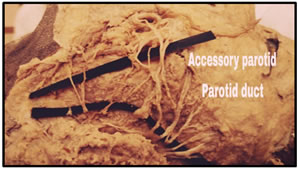
CASE REPORT During routine cadaveric dissection, in a 45 year old male cadaver, we found the presence of extra gland mass anterior to the stenson’s duct in parotid gland dissection. This mass was approximately 1X1 cm in dimension. It was located in the vicinity of parotid gland on the main parotid duct. On further meticulus dissection, no separate duct was found for this accessory gland mass. As it was located on the main Stenson’s duct, there is possibility of emptying of secretions of this accessory gland directly into the main duct without any secondary duct like in common scenarios of accessory parotid. This accessory parotid was covered by network of intercommunications between the different branches of facial nerve forming parotid plexus. The intercommunications were present in the form of loop formed mainly between the zygomatic and upper buccal branches and joined by marginal mandibular branch.
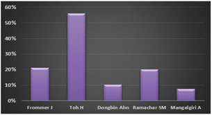
DISCUSSION Accessory salivary gland is a normal variant of the collection of salivary tissue away from the main parotid gland. They are normally located on lateral aspect of the masseter muscle. The knowledge of accessory parotid is necessary as any pathology of main parotid gland involves accessory parotid gland. This can be attributed to histological similarity between the two.9 Ammar Haouimi mentioned in his article that an accessory parotid gland has the same MRI signal and CT density as that of the main parotid gland. When compared, the aging changes in the main parotid gland like increased connective tissue and fat and decreased glandular elements are more extensive compared to those of accessory parotid gland.4 The varied incidence rate of accessory parotid gland is found in different studies in the literature. When reviewed, highest rate found was 56% in the study by Toh H07 while the low incidence was reported by author Mangalgiri A in his study which was 7.5% Table 1: showing incidence rate of accessory parotid gland in different studies
Graph 1: showing incidence rate of accessory parotid gland in different studies
Figure: showing accessory parotid gland located anterior to the main parotid duct and covered by intercommunications between different branches of facial nerve The presence of accessory parotid gland can complicate parotid surgeries and can serve as potential site of benign and malignant tumor of parotid gland.10 Accessory parotid gland is the site for congenital and acquired lesions. It is the commonest site for pleomorphic adenoma. The incidence of accessory parotid gland tumors is more than the main gland.9 Travis R11 in his study mentioned that out of 152 cases of accessory parotid gland lesions 70% were benign and 30% were malignant. The author Zhu W12 reported the incidence of accessory parotid gland was 71.8% in all cases of parotitis studied. This rate is higher than the rate in healthy subjects. This finding was in agreement with the finding of author Horsburgh13 who reported the incidence of 59%. As per the author, the main pathogenesis for parotitis is reduced salivary flow which can be more due to sialolith or mucous emobolus in accessory parotid gland caused by extra gland mass and confluence. Accessory parotid gland is normally mid cheek swelling. Surgical resection is the first choice of treatment for accessory parotid gland tumors.14 As emerging branches of facial nerve from temporofacial and cervicofacial trunks are located at this site, the surgical removal of gland can damage these terminal branches leading to the facial paralysis.10 So the surgeons must consider the possibility of presence of accessory parotid gland during parotidectomy to avoid post-operative complications.
CONCLUSION Presence of accessory parotid gland is potential risk factor for parotitis and benign and malignant tumors of parotid. Any pathology involving parotid gland also involves accessory parotid gland so it is imperative for surgeons to have knowledge of location and possible implication of accessory parotid while dealing with clinical conditions associated with parotid.
REFERENCES
Policy for Articles with Open Access: Authors who publish with MedPulse International Journal of Anatomy (Print ISSN: 2550-7621) (Online ISSN: 2636-4557) agree to the following terms: Authors retain copyright and grant the journal right of first publication with the work simultaneously licensed under a Creative Commons Attribution License that allows others to share the work with an acknowledgement of the work's authorship and initial publication in this journal. Authors are permitted and encouraged to post links to their work online (e.g., in institutional repositories or on their website) prior to and during the submission process, as it can lead to productive exchanges, as well as earlier and greater citation of published work.  This work is licensed under a Creative Commons Attribution 4.0 International License. |
|
 This work is licensed under a Creative Commons Attribution-NonCommercial 4.0 International License.