 Home
Home
|
Table of Content Volume 17 Issue 1 - January 2021
Anomalous ganglion associated with varied terminal branching pattern of facial nerve
Alka Bhingardeo
Assistant Professor, Department of Anatomy, All India Institute of Medical Sciences, Bibinagar, INDIA. Email: dralkabhingardeo@gmail.com
Abstract Background: Facial nerve is the mixed cranial nerve. It is the nerve of second pharyngeal arch. It gives five terminal branches after emerging from the parotid gland which supplies different muscles of facial expression. These are one each temporal, zygomatic, buccal, marginal mandibular and cervical branch. Buccal branch further divides into upper and lower buccal branches. During routine cadaveric dissection we found bilateral varied terminal branching pattern of facial nerve which showed four temporal, two marginal mandibular and two cervical branches with two loops and two H shaped intercommunications. An anomalous ganglion is found in association with a these teminal branches which had given a communicating branch to the loop formed by zygomatic, buccal and marginal mandibular branch. To the best of my knowlege presence of such anomalous ganglia associated with terminal branches of facial nerve is not mentioned anywhere in the literature. We also found an accessory parotid gland located near the main parotid duct without secondary duct. The functional conservation of facial nerve branches in surgeries around parotid region and procedures like facial rhytidectomy is necessary. To avoid all the postoperative morbidity associated with the injury to the facial nerve, detailed knowledge of the branching pattern of the facial nerve and its probable variations is must. Key words: facial nerve, anomalous ganglion, parotid surgery, facial nerve paralysis, accessory parotid gland
INTRODUCTION Facial nerve is also called as ‘Smiling nerve’ as it supplies all the muscles of facial expression. It is the seventh cranial nerve which emerges from the surface of pons. It enters the parotid gland through it’s posterior border and passes through superficial and deep part of the gland winding around isthmus. It divides into two trunks inside the gland, these are - temporofacial and cervicofacial. Temporofacial trunk further gives rise to temporal and zygomatic branch while cervicofacial trunk divides into buccal, marginal mandibular and cervical branch. Buccal branch further divides into upper and lower buccal branches. Normally one each temporal, zygomatic, buccal, marginal mandibular and cervical branch are present1,2. Variations in terminal branching pattern of facial nerve are not uncommon. Devis classified branching pattern into six different categories depending upon communications present between the branches while Kwak HH classified it into five categories depending upon the origin of buccal branch.
CASE REPORT During routine cadaveric dissection, in an adult male cadaver, we found bilateral varied pattern of terminal branching pattern of facial nerve. Variations in the branches of temporofacial trunk -
Variations in the branches of cervicofacial trunk -
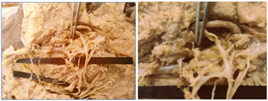
An anomalous ganglion was found among these nerve twigs. Near the loop formed by zygomatic, buccal and marginal mandibular branch an anomalous ganglion was present. There were many small nerve twigs arisinf grom this ganglion supplying surrounding structures. It also gave one communicating branch to the loop. The presence of such anomalous ganglia at the terminal branch of facial nerve is not reported anywhere in the literature. To the best of my knowledge, this is the first case of such anomalous ganglia. We also found an accessory parotid gland approximately 1cm X 1cm size located in anterior to the main parotid duct. On further dissection, no secondary duct was found for this accessory parotid gland.
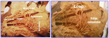
Figure 1 Figure 2 Figure 3 Figure 4 Figure 1: showing different terminal branches of facial nerve [MM -Marginal mandibular]; Figure 2: showing different intercommunications between terminal branches of facial nerve; Figure 3: showing an anomalous ganglion associated with nerv e twigs; Figure 4: showing closer view of an anomalous ganglion with emerging rami
DISCUSSION The variations in the terminal branches of facial nerve is not uncommon. In our case, we found variations in the number of branches of temporofacial and cervicofacial trunk and intercommunications between the different branches. We found that temporal branch was represented by total four rami. Gosain A K3, mentioned in his article that as per recent studies temporal branch is not a single branch but represented by more than one branches.We also found communicating branch between zygomatic and adjascent temporal branch giving H shape to the communication.We found total three ramifications of zygomatic branch. Out of these two branches had the same course and supplied same group of muscles while one branch deviated from the course and joined the upper buccal branch for a short distance and then got separated and formed a loop with the upper buccal branch. A communicating branch is also found between the zygomatic and buccal branch at the level of emergence from the anterior border giving H-shape to the communication and two branches. The communication between zygomatic and buccal branch is the communication between cervicofacial and temporofacial trunk. Author Gosain A K3 in his study found such interconnections between the branches of two trunks in more than 70% of cases while Bandell H4 found in 44% of cases. In case of cervicofacial trunk, we found a single buccal branch which further divided into upper and lower buccal branch. There was a loop of coomunication between the upper and lower buccal branches which is joined by twig from the marginal mandibular branch. Sometimes rather than one buccal branch dividing into upper and lower buccal we get two buccal branches separately. Liu AT5 in his study on facial nerve found two buccal branches in 87.5% of cases. We found two ramifications of marginal mandibular nerve both of which joined lower buccal branch and formed a loop. Author Karapinar U6 et al. in his study found such two ramifications of marginal mandibular nerve in 63.6% of cases. The author in his article also mentioned communication between marginal mandibular and buccal branches in 4.6% of cases but had not mentioned the structure or type of communication. In case of cervical banch, we found immediately after arising from the trunk, the cervical branch divided into two branches out of which the upper branch again ramified into two rami. One of the ramus from the upper cervical branch formed a loop with marginal mandibular branch. Our this finding of communication between marginal mandibular branch not only with buccal but also with the cervical branch is similar to the findings by author Woltmann M7. The interconnections between the different branches of temporofacial and cervicofacial trunk or within the branches of same trunk are important. There is increased risk of facial paralysis in case the terminal branch is transected if the interconnections between the branches of facial nerve are less1. In procedures like facial rhytidectomy, coronal or endoscopic bro lifting and temporal craniotomy there is more probability of injury to temporal and zygomatic branch while in surgeries of submandibular region there are chances of injury to marginal mandibular branch8. Saylam C9 in his article mentioned that marginal mandibular branch is more prone to injury in procedure of rhytidectomies and surgeons must be aware of probable variations in location and branching pattern of marginal mandibular branch.When compared, our case can be included in sixth category of Devis classification where multiple anastomosis are found between temporofacial and cervicofacial divisions and marginal mandibular branch send twigs to temporofacial branch. In case of Kwak’s classification, it can be included into category four where communication is present between nerve twigs from the zygomatic and marginal mandibular branches with buccal branches. A unique anomalous ganglion of pin head size was found near the loop formed by zygomatic, buccal and marginal mandibular branch. So many rami were seen emerging from this ganglion. One of the ramus supplied a communicating branch to the adjascent loop. Among the different extensions of parotid gland the anterior extensions are two - one is ‘Facial process’ which is attached directly to the gland and another is ‘accessory parotid gland’ which is completely separated from the main gland like a detached glandular mass10. An accessory parotid gland is the salivary tissue present in close association or anterior to the Stensen's duct, lying on the masseter muscle but away from the main parotid gland11,12. It is found in 21-61% of cases as per different autopsy studies11,13. In our case, we found an accessory parotid gland which was located anterior to parotid duct or stensen’s duct and was approximately 1X2 cm in size without any secondary duct.
CONCLUSION To avoid all the postoperative morbidity associated with the injury to the facial nerve, detailed knowledge of the branching pattern of the facial nerve and its probable variations is must. As accessory parotid gland shows all the tumor types which occur in the main parotid gland and recurrence rate of such tumors is high, it is imperative for surgeons to have knowledge of accessory parotid gland and its related variations.
REFERENCES
Policy for Articles with Open Access: Authors who publish with MedPulse International Journal of Community Medicine (Print ISSN: 2579-0862) (Online ISSN: 2636-4743) agree to the following terms: Authors retain copyright and grant the journal right of first publication with the work simultaneously licensed under a Creative Commons Attribution License that allows others to share the work with an acknowledgement of the work's authorship and initial publication in this journal. Authors are permitted and encouraged to post links to their work online (e.g., in institutional repositories or on their website) prior to and during the submission process, as it can lead to productive exchanges, as well as earlier and greater citation of published work.
|
|
 This work is licensed under a Creative Commons Attribution-NonCommercial 4.0 International License.