 Home
Home
|
Table of Content Volume 17 Issue 3 - March 2021
A bilateral variant accessory belly of extensor indicis muscle in posterior compartment of forearm: An anatomical variation
Sushma Daripelli1*, Mrudula C2, Alka B3, Surraj S3
1Senior Resident, 2Additional Professor, 3Assistant Professor, Department of Anatomy, All India Institute of Medical Sciences, Bibinagar, Telangana, India 508126, INDIA. Email: sushma.tulips@gmail.com
Abstract Background: Anomalous extensor muscles of the hand are not uncommon. There are numerous reports regarding the variations of the extensor muscles generally encountered during surgical and dissection procedures. Objectives: variations in the muscles of external compartment of forearm are important in interpreting rare clinical conditions like Extensor Indicis Proprius Syndrome. Knowledge of these kinds of variation are useful for Hand surgeons to plan surgical procedures during muscle graft and tendon transplantation procedures. Material and Methods: The present study was conducted in 10 embalmed upper limb specimens obtained from adult cadavers collected from Department of Anatomy, AIIMS Bibinagar Telangana, India. Result: The present case report is on the unusual bilateral variant Accessory Belly of extensor indicis muscle. Both the additional bellies of right and left forearm originated from the main belly of Extensor Indicis, presented with two tendon slips, passing through the fourth compartment of the extensor retinaculum, on the right forearm one of these tendinous slips is inserted into ulnar aspect of the dorsal digital expansion of the middle finger, the other slip was merged into the dorsum of the hand. Both additional belly’s was supplied by posterior interosseous nerve. on the left forearm one of the tendinous slip is inserted into the dorsal part of capsule of the metacarpophalangeal joint of the middle finger and the other tendinous slip merged into the dorsal digital expansion of middle finger. Conclusion: clinicians must be conscious of these variations for successful diagnosis and treatment of Extensor Indicis Proprius Syndrome and tendon grafting. Key Words: Accessory Belly, Extensor indicis tendon, Tendon Transplantation, Extensor proprius syndrome.
INTRODUCTION The Extensor Indicis(EI) muscle is one of the known deep extensor of forearm, which lies medial and parallel to extensor pollicis longus.1 It helps in extension of index finger and wrist. Tendons may be single or double or triple at the myotendinous junction. Tendon slips are defined as division of tendon or splitting of tendon into two or more slips2. Tendon injuries in dorsum of hand are common. Awareness on these kinds of variations are important for Hand surgeons to plan surgical procedures during muscle graft and tendon transplantation procedures and for diagnosis of clinical syndromes like Extensor Indicis Proprius Syndrome.
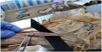
CASE DESCRIPTION During routine dissection of teaching for phase I undergraduate medical students in AIIMS Bibinagar, we found an anomalous additional belly of Extensor Indicis(EI) muscle bilaterally in an adult male cadaver of around 70 years of age. The EI muscle originated as a small belly from the posterior surface of the ulna and adjoining interosseous membrane, passes through the fourth compartment deep to extensor retinaculum and inserted into the ulnar aspect of the extensor expansion of the index finger opposite the head of second metacarpal bone. In the present case, we observed an unusual Configuration of additional Belly of extensor indicis muscle and its tendinous slips were noted in both upper limbs. Both the additional bellies of right and left forearm originated from the main belly of EI, presented with two tendon slips passing through the fourth compartment of the extensor retinaculum. Both additional bellies were supplied by posterior interosseous nerve. on the right forearm one of these tendinous slips is inserted into ulnar aspect of the dorsal digital expansion of the middle finger, the other slip was merged into the dorsum of the hand (Figure 1). on the left forearm one of the tendinous slip is inserted into the dorsal part of capsule of the metacarpophalangeal joint of the middle finger and the other tendinous slip merged into the dorsal digital expansion of middle finger (Figure 2).
Figure 1: 1: photograph showing the Additional Belly of extensor indicis muscle and its insertion on right sided forearm. (EI: extensor indicis, *: Additional belly , ED: extensor digitorum tendon).
Figure 2: photograph showing the Additional Belly of extensor indicis muscle and its insertion on Left sided forearm. (EI: extensor indicis, *: Additional belly ,ED: extensor digitorum tendon.)
DISCUSSION The extensors of forearm are known to exhibit wide range of variations. Among the extensors, the EI is well known to associate with the variation of its muscle belly and also with tendon and its insertions. The EI is widely used in surgeries for tendon transfer, designed to restore finger movements because index finger receives extensor digitorum tendon3. It is utilized in tendon graft surgeries in functional loss of extensor pollicis longus, abductor pollicis muscle, and opponens pollicis muscle4,5. In the present case, we noticed unique and rare variation of EI, in the right upper limb the accessory muscle belly was divided into two tendinous slips, the uniqueness is in this insertion of One of the slip is attached to ulnar aspect of the dorsal digital expansion of the Middle Finger, the other slip was merged into the dorsum of the hand. In the left upper limb accessory muscle belly was divided into two tendinous slips, the uniqueness is in this insertion of one tendinous slip into the dorsal part of capsule of the metacarpophalangeal joint of the middle finger and the other tendinous slip merged into the dorsal digital expansion of middle finger. In a study by yoshida et al., the additional EI had two slips, both were attached to the tendon of Extensor Digitorum of the middle finger. Srinivasa et al.6 reported additional EI had two tendon slips, one slip is attached to the ulnar side of Extensor Digitorum tendon of the middle finger and the other slip is merged with the fascia over dorsum of the hand. EI was absent in both hands of cadaver was reported by zilber and oberlin et al.7 The present case is in contradiction to the above reported cases, it demonstrates additional belly of EI with two tendons with two different sites of insertion. Knowledge about the presence of multiple tendons of extensors of forearm are useful in identifying and planning tendon transfer or graft surgeries. The additional bellies can be used for the muscle graft, if the muscle belly are small they get unnoticed, if muscle belly is large it confuse surgeons during surgeries operating in the forearm extensor and hand regions and clinicians during diagnosis of soft tissue conditions. Some times when the additional tendons are thick, it decreases space in the respective compartment it results the risk of tenosynovitis. Insertion of additional tendon on the capsule may results in ganglion formation or may restricts the movement of joint, which is seen in this case. Extension of a musculotendinous portion of EI into the fourth dorsal compartment of wrist leads to Extensor Proprious Syndrome8, which is characterised by dorsal wrist pain and synovitis of EI muscle within the fourth dorsal compartment of wrist, aggravated by activities. Anatomical knowledge of variations in extensor tendons is of clinical importance in dorsum of hand. Accessory tendons if present can be utilized for tendon graft rather than utilizing from distant site, by routine radiological assessment of extensor tendons, its variations is useful during tendon graft surgeries. The present case report with its uniqueness and rarity adds knowledge to the surgeons and clinicians in efficacious treatment and diagnosis.
CONCLUSION precise knowledge and awareness of variations of extensor indicis is useful for clinicians to understand better diagnosis and treatment and helps in assessing, repair and treating hand injuries and disorders. ACKNOWLEDGEMENTS: I would like to thank those who have donated their bodies and their families to anatomical research. No funding was provided.
REFERENCES
Policy for Articles with Open Access: Authors who publish with MedPulse International Journal of Community Medicine (Print ISSN: 2579-0862) (Online ISSN: 2636-4743) agree to the following terms: Authors retain copyright and grant the journal right of first publication with the work simultaneously licensed under a Creative Commons Attribution License that allows others to share the work with an acknowledgement of the work's authorship and initial publication in this journal. Authors are permitted and encouraged to post links to their work online (e.g., in institutional repositories or on their website) prior to and during the submission process, as it can lead to productive exchanges, as well as earlier and greater citation of published work.
|
|

 This work is licensed under a Creative Commons Attribution-NonCommercial 4.0 International License.