 Home
Home
|
Table of Content Volume 17 Issue 3 - March 2021
Histochemical study of mucosubstances in normal human vermiform appendix
Avinash D Shewale1, Swati Lalasaheb Patil2*, Rohini R Karambelkar3
1Associate Professor, Department of Anatomy, BKLWRMC Sawarde, Chiplun, Maharashtra, INDIA. 2Associate Professor, 3Professor and HOD, Department of Anatomy, PIMS and R Islampur, Sangli, Maharashtra, INDIA. Email: drswatipatil1612@gmail.com
Abstract Background: The gastro intestinal tract is lined by epithelium, secreting mucosubstances which perform a wide variety of functions like lubrication, protection against acids etc. In present study mucosubstances present in the lining epithelium and crypts of human vermiform appendix are studied. Twenty normal human appendices obtained during autopsies or surgical removal were collected and fixed in 10% formalin with 2% calcium acetate and a pinch of phosphotungstic acid. 4 to 5 micron thin sections were cut after preparing paraffin blocks. The sections were stained by using specific stains like periodic acid schiff, Alcian blue of different pH and Aldehyde fuschin singly and in combinations. Confirmatory tests were also carried on. The normal human vermiform appendix shows presence of acidic mucosubstances mainly, which are predominantly carboxylated or weakly acidic. The carboxylated mucosubstances possess an antiviral and antibacterial property. Any change in its nature makes vermiform appendix vulnerable for infection. Key Words: Mucosubstances, Vermiform Appendix, PAS, AB, AF.
INTRODUCTION The gastrointestinal tract is lined by epithelium, secreting mucosubstances which perform a wide variety of functions like lubrication, protection against acids etc. The mucosubstances also contain immunoglobulin, principally of IgA type, Lactoferrins which chelate the iron necessary for growth of some bacteria and lysozymes which destroy some of bacteria.1 The human vermiform appendix was previously considered as vestigial organ, but now it is known that it contains a ring of lymphoid tissue and is important immunologically. Hence it is considered as abdominal tonsil. Many workers like Dr.Lala (1987)2 and Dr. Ganga (2004)3 have studied mucosubstances in large intestine and other parts of gastrointestinal tract but epithelial mucins in vermiform appendix have remained a bit neglected and hence the present study was undertaken.
MATERIAL AND METHODS Ten normal human vermiform appendices obtained during autopsies or surgical removals were collected. The tissue was fixed in 2% calcium acetate in 10% formalin with a pinch of phosphotungstic acid for two days. By routine procedure paraffin blocks were prepared and 4-5 micron thin sections were cut. They were stained by Haematoxylene – Eosin. Periodic acid schiff reagent. PAS Diastase 4. PAS Phenyl hydrazine. Alcian Blue 2.5pH. Alcian Blue 1pH. Combined AB-PAS. Aldehyde fuschin. Combined AF-AB 5.
OBSERVATIONS AND RESULTS The vermiform appendix being a part of large intestine is lined by simple columnar epithelium with plenty of goblet cells as seen in high power view of the slide stained with H. E. (Photo I)
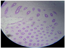
Photo I: Showing H and E staining (High Power) The epithelium lining the surface and crypt stained intensely with P.A.S. showing magenta colour. (Photo II)
Photo II: PAS staining The intensity remains unaffected by diastase digestion (photo III).
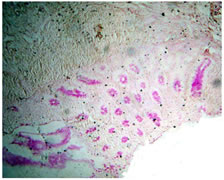
Photo III: PAS Diastase Staining No change in intensity even with Phenyl hydrazine (photo IV) indicates the presence of acidic mucosubstances.
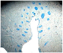
Photo IV: Phenyl Hydrazine staining. Blue staining showing positive results with AB 2.5 Ph (photo V) are seen.
Photo V: AB 2.5pH staining. Very faint or negative results are seen with AB Ph 1 (photo VI).
Photo VI: AB 1pH staining. Similarly negative results with AF (photo VII ) indicated that the mucosubstances are acidic and predominantly carboxylated one.
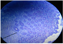
Photo VII: AF staining. Mainly Blue colour with no magenta or purple colour is seen in combined AB-PAS (photo VIII).
Photo VIII: AB-PAS staining Mainly blue colour seen in AF AB (photo IX) staining confirmed the presence of carboxylated or weak acidic mucosubstances.
Photo IX: AF AB staining. The nomenclature applied to the mucosubstances is taken from the discussion of a proposed terminology of histochemistry recognizable material 6. The observations are tabulated according to visually estimated intensity of staining and shade as in table I.
Table 1: SHOWING COLOUR INTENSITY OF STAINS USED
DISCUSSION The mucosubstances have been classified into neutral and acidic types, the later being further subdivided into Sulphated (strong acidic) and nonsulphated ( carboxylated or weak acidic) 6. The epithelial mucosubstances found in human vermiform appendix are predominantly carboxylated or weak acidic. Carboxylated mucosubstances are rich in lysozymes and possess antibacterial and antiviral property.7 Any reduction or chemical change in the nature of mucosubstances may make vermiform appendix more vulnerable for infection. This will be understood better only when further studies will be carried out in various diseases of appendix and compared with normal one.
CONCLUSION The human vermiform appendix was previously considered as a vestigial organ but now it is observed that it secretes carboxylated mucosubstances and provides immunity against various infections.
ACKNOWLEDGMENT We are very much thankful to department of surgery, pathology and our lab technician for helping us in compilation of this research work.
REFERENCES
Policy for Articles with Open Access: Authors who publish with MedPulse International Journal of Community Medicine (Print ISSN: 2579-0862) (Online ISSN: 2636-4743) agree to the following terms: Authors retain copyright and grant the journal right of first publication with the work simultaneously licensed under a Creative Commons Attribution License that allows others to share the work with an acknowledgement of the work's authorship and initial publication in this journal. Authors are permitted and encouraged to post links to their work online (e.g., in institutional repositories or on their website) prior to and during the submission process, as it can lead to productive exchanges, as well as earlier and greater citation of published work.
|
|


 This work is licensed under a Creative Commons Attribution-NonCommercial 4.0 International License.