 Home
Home
|
Table of Content - Volume 18 Issue 2 - May 2021
Study on variations in hilar and segmental branching pattern of splenic artery
Swapnilkumar L Sarda1*, M S Selukar2, P R Chavan3, P R Kulkarni4
1Assistant Professor, Department of Anatomy, Shri. Bhausaheb Hire Government Medical College, Dhule, INDIA. 2Associate Professor, Department of Anatomy, Government Medical College Latur, INDIA. 3Assistant Professor, Department of Anatomy, Government Medical College, Aurangabad, INDIA. 4Professor and Head, Department of Anatomy Ashwini Rural Medical College Solapur, INDIA. Email: drswapnilsarda@yahoo.com
Abstract Background: Introduction of laparoscopic surgical methods requires exact knowledge of the topography of the spleen and its surrounding. Further advances in splenic conservative surgery are dependent on better knowledge of vascular anatomy of the spleen. Hence segmental arteries of spleen are of great surgical importance and their early identification in splenic trauma will lead to enhanced splenic conservation. Materials and methods: Present study carried out on 50 human spleen by dissection, silicon injection and radiological methods. We found different types of variations in hilar and segmental branching pattern of splenic artery. Observations and Results: in present study splenic artery divided in two primary branches in 86% and in three primary branches in14% of the spleens. superior polar branches were found in 44% while inferior polar branches were in 52% of the spleens. Two segmental branches found in 24%, three segmental found in 50%, four segmental in 18% and five segmental in 8% of the specimens. Intersegmental anastomosis found in 2% of spleens. Discussion: These segmental resection of spleen and further advances in splenic conservation are dependant on better understanding of vascular anatomy of the spleen. Keywords: Splenic artery, hilar branchs, segmental arteries
INTRODUCTION The human spleen is an organ demanding constant attention from anatomical, immunological, surgical and clinical point of view. The spleen shows segmentation due to fibrous septa described by Kyber1 (1870) in man, cat, dog, horse and rabbit . Partial splenectomy was first performed in 19th century and its post operative sequel and complications were observed (Greco and Alvarez, 1981)2. Thus the application of conservative surgery of partial splenectomy became a worldwide practice. Gupta et al.3 (1976) reported the avascular plane and the segmental pattern of the spleen like those in other species. Each segment is having hilar branch of the main splenic artery and splenic vein. The application of the conservative splenic surgery requires a detailed knowledge of the avascular plane of the spleen and its segmental pattern in both male and female (Chakravarthy S, 2003)4. The splenic artery divides into two or three primary or hilar branches [terminal branches]. The arteries supplying one of the poles of the spleen are polar arteries (Michel NA)5. A vessel is considered as polar artery, when it penetrates one of the splenic poles and not the hilum. There is presence of superior polar branch [which supply posterior pole / extremity] or inferior polar branch [which supply anterior pole / extremity] or both; superior and inferior polar branches. These polar branches may originate from primary branches of splenic artery or from the trunk of splenic artery itself. The human spleen is divided accordingly into two or three primary segments, separated by a definite avascular plane. Also, a rather constant avascular plane separates the polar segments from remaining of the organ. Thus, the number of segments of spleen, may vary from two to five. The arteries supplying these segments are segmental arteries.
MATERIALS AND METHODS Total Fifty human spleens out of that fourty nine from the embalmed cadavers from Department of Anatomy and one postmortem specimen of human spleen from Department of Forensic, of various medical colleges were obtained for the study. The spleens were stored in 40% formalin solution in different jars and numbered serially.
The spleen was removed along with long splenic artery from origin. The soft tissue (fat) attached to hilum was removed to expose the branches of splenic artery. After that each spleen was dissected carefully by piece-meal dissection method. The splenic artery and its branches were cleaned and traced. Segmental branches of splenic artery were identified and traced individually. The pattern of branching was seen for each spleen. Any variation in form of number of segmental branches and intersegmental anastomosis if present, was noted and photographed.
Spleens were collected from the Anatomy department. The splenic artery was dissected and warm saline was injected into the artery. The spleen was then placed for about an hour with the artery turned downwards to drain out the fluid. Then the silicon gum, loaded in 5ml syringe with 16 bore needle, was injected into the splenic artery. The specimen was kept for 48 hours to allow the silicon gum to settle. Then it was boiled in hot water bath for 15-25 minutes to take off the splenic tissue. The resulting cast was cleaned carefully and then dried. Hilar branches and segmental branches were observed, specimen was numbered and photograph taken.
Fresh spleen was collected from the department of Forensic. The splenic artery was dissected and warm saline was injected into the artery. The spleen was then placed for about an hour with the artery turned downwards to drain out the fluid.Then Barium sulfate-- liquid contrast solution was injected into the splenic artery. The spleen was kept overnight in refrigerator. On next day radiographs were taken. The radiographs were studied and the hilar branches and segmental branches of splenic artery were noted down. OBSERVATIONS AND RESULTS In the present study, fifty human spleens were observed to study hilar and segmental branches of splenic artery.
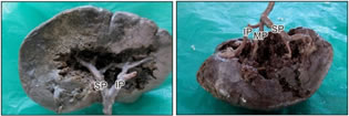
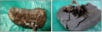
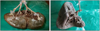
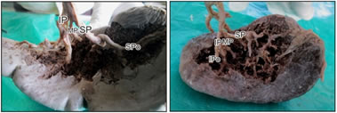
Photograph 1 Photograph 2 Photograph 3 Photograph 4 Photograph 5 Photograph 6 Photograph 7 Photograph 8 Photograph 1: Two primary branches,[ SP - Superior Primary, IP – Inferior Primary]; Photograph 2: Three primary branches, [SP –Superior Primary, MP – Middle Primary, IP – Inferior Primary]; Photograph 3: Superior polar branch arising from splenic trunk, [SPo – Superior Polar ]; Photograph 4: Superior polar branch arising from superior primary branch; Photograph 5: Inferior polar branch arising from splenic trunk[ IPo – Inferior Polar]; Photograph 6: Inferior polar branch arising from inferior primary branch; Photograph 7: Two primary and two polar branches; Photograph 8: Three primary and two polar branches; Photograph 9: Three primary and one superior polar; Photograph 10: Three primary and one inferior polar Photograph 11: Vascular anastomosis betn segmental branches; Photograph 12: Silicon injection cast; Photograph13: Radiological study
Table 1: Primary or Hilar division of splenic artery
Table 2: Frequency of polar arteries
Table 3: Origins Of Superior Polar Artery
Table 4: Origin of inferior polar artery
Table 5: Association between polar branches and primary / Hilar branches
From table no.1 and 5, it is clear that-
Out of 43 spleens–
Out of those 07 spleens –
From above observations, it is clear that the number of segmental branches varied from two to five. The segmental branches were as follows:
Interarterial anastomosis In spleen no. 7, splenic artery divides into three primary branches superior, middle and inferior. It is observed that there is vascular anastomosis between superior and middle primary / hilar branches of splenic artery. [Photograph no.11] So in only one spleen [2%] interarterial anastomosis was present.
DISCUSSION As reported by Michel NA5 (1942), Gupta et al.3 (1976), Mikhail et al.6 (1979) Garcia et al.7 (1988) and many other research workers and as mentioned in Gray’s anatomy (39th edition 2005), in the present study also, many variations were found in the polar and segmental branches of splenic artery. Two primary branches; superior and inferior were observed in 86% specimens and three primary branches; superior, middle and inferior were observed in 14% specimens in present study. These observations are in agreement with previous studies . Michel NA5 had found 80% two and 20% three primary branches. Gupta et al.3 had found 84% two and 16% three primary branches. Mikhail et al.6 had found 77% two and 23% three primary branches. Katritsis et al.8 had found 85.70% two and 14.30% three primary branches. Chaware et al.9 had found 85.58% two and 14.42% three primary branches. Superior polar arteries were found in 44% and inferior polar arteries were found in 52% spleen. These finding also are in agreement with findings of previous studies. Michel NA5 had found 65% superior polar and 82% inferior polar arteries. Katritsis et al.8 had found 60% superior and 80% inferior polar arteries. Chaware et al.9 had found 40.53% superior and 54.06% inferior polar arteries. The origin of polar arteries was variable. Superior polar artery originated from superior primary branch in 86.37% spleens and from splenic trunk in 13.63% spleens. Inferior polar artery originated from inferior primary branch in 76.93 % and from splenic trunk in 23.07% spleens. These findings were in accordance with Michel NA5 (1942) and Garcia and Lemes7 (1988). They reported origin of polar arteries from superior and inferior primary branches and from splenic trunk. They also reported rare origin of arteries from left gastric artery and from short gastric artery, which was not observed in the present stud In the present study two segmental branches were present in 24% spleens, three segmental branches in 50% spleens, four segmental branches in 18% spleens, five segmental branches in 08% spleens. These observations are not in relation with previous studies as . Mandarim et al.10 had found that two segmental branches were present in 68.2% spleens, three segmental branches in 10.6% spleens, four segmental branches in 4.5% spleens, five segmental branches in 6.7% spleens. Chaware et al.9 had found that two segmental branches were present in 13.51% spleens, three segmental branches in 60.66% spleens, four segmental branches in 17.11% spleens, five segmental branches in 2.7% spleens. Thus no. of segmental arteries are highly variable. Variations in segmental branches of splenic artery depends upon its development. Spleen appears at first as a number of lobules in the dorsal mesogastrium. These lobules join together to form a single splenic mass. These embryological facts points out variations in segmental branches of splenic artery. In most of the studies carried out earlier, only in three studies there was vascular anastomosis between adjacent segments of spleen. In the present study, intersegmental anastomosis was found in 2% of the total spleens. Mandarim et al.10 had found 16.7%, Garcia et al.7 had found 19.8%, Chaware et al.9 had found that 1.80% spleens.
CONCLUSIONS In the present study, splenic artery divided into two primary branches in 86% and into three primary branches in 14% of the spleens. Superior polar branches were found in 44% while inferior polar branches were in 52% of the spleens. The primary branches and polar branches of splenic artery which supplied the corresponding lobes of the spleen and divided spleen into segments, separated by a definite avascular plane, are called as segmental branches. In the present study number of segmental branches varied between two to five. Two segmental branches were found in 24%, three segmental branches in 50%, four segmental branches in 18% and five segmental branches in 8% of the specimens. In the present study intersegmental anastomosis was found in 2% of the spleens.
REFERENCES
Policy for Articles with Open Access: Authors who publish with MedPulse International Journal of Pediatrics (Print ISSN: 2579-0897) (Online ISSN: 2636-4662) agree to the following terms: Authors retain copyright and grant the journal right of first publication with the work simultaneously licensed under a Creative Commons Attribution License that allows others to share the work with an acknowledgement of the work's authorship and initial publication in this journal. Authors are permitted and encouraged to post links to their work online (e.g., in institutional repositories or on their website) prior to and during the submission process, as it can lead to productive exchanges, as well as earlier and greater citation of published work.
|
|
||||||||||||||||||||||||||||||||||||||||||||||||||||||||||||||||||||||||||||||||


 This work is licensed under a Creative Commons Attribution-NonCommercial 4.0 International License.