 Home
Home
|
Table of Content - Volume 19 Issue 1 - July 2021
Assessment of DNA damage through micronucleus studies in subjects exposed to formalin
K Sriambika1*, Magi murugan2, Rema Devi3
1Assistant Professor, 2Professor, 3Professor & HOD, Department of Anatomy, Pondicherry Institute of Medical Sciences, Ganapathy Chettykulam, 605014, Pondicherry, INDIA. Email: kambika1784@gmail.com
Abstract Background: Formaldehyde (FA) is the reactive and simplest of all the aldehydes. It is used as a preservative in anatomy, pathology and forensic laboratories. The international agency for research on cancer has classified FA as a carcinogen that can cause nasopharyngeal carcinoma, Leukaemia, Liver and pancreatic cancer. Objective And Method: The aim of the study was to assess the DNA damage in peripheral blood lymphocytes and in buccal cells by Micronucleus assay in Formalin exposed workers of Anatomy, Pathology and Forensic laboratories and compare with the control group, and also to analyze the relationship between frequency of Micronuclei and duration of exposure to formalin. Results: The mean and standard deviation (SD) of micronuclei in peripheral blood of exposed was 8.35 and in controls was 4.18. There was a significant increase in the frequency of MN in exposed group when compared with the comparison group (p<0. 5876). Pearson’s correlation test showed a positive correlation between the years of FA exposure and the number of micronuclei in buccal cells and peripheral blood indicating that DNA damage due to FA was directly proportional to the duration of exposure (r=0.8, 0.9). Conclusion: The present study was done to assess the DNA damage in people who were exposed to FA and a control group not exposed to FA by buccal cell and peripheral blood Micronucleus Assay. There was a significant increase in the MN in people exposed to FA which was directly proportional to the duration of exposure. Keywords: DNA damage, buccal cells, Formaldehyde, Peripheral blood, Micronuclei.
INTRODUCTION Formaldehyde (FA) is the reactive and simplest of all the aldehydes. It usually occurs in gaseous form in nature. Because of its high-water solubility, the aqueous solution is used in Anatomy, Forensic and Pathology departments as a preservative and fixative.4 It occurs as a natural product in most organisms due to various metabolic processes. Absorption of formalin is through inhalation and skin where it acts as an irritant and allergen and thereby causes asthma, dermatitis, and irritation in the conjunctiva as well as mucosa of respiratory tract. The international agency for research on cancer has classified FA as a carcinogen that can cause nasopharyngeal carcinoma, Leukaemia, Liver and pancreatic cancer.1 Various indicators are used to detect the cytogenetic alterations caused due to FA exposure. Among them Micronucleus test is a sensitive and well-established tool for measuring the DNA damage.4 The current study aims at detecting the DNA damage in peripheral blood and buccal mucosa cells by using the cytokinesis blocked micronucleus assay (CBMA), which is one of the most reliable method for detecting and evaluating DNA damage. This study was taken to highlight the effect of formalin in workers exposed to formalin in anatomy, pathology and forensic laboratories. Since the exposure to formaldehyde causes adverse effects like genotoxicity, carcinogenicity and teratogenicity this study points the need for the close monitoring of occupational exposure to FA and to reduce the risk by using appropriate protective measures. OBJECTIVES
METHODOLOGY The present cross-sectional study was carried out in the Division of Human Genetics, Pondicherry Institute of Medical Sciences. The sample size was calculated using OpenEpi software version 3.03. Ethical clearance was obtained from the Institutional Ethics committee. 28 subjects from Anatomy, Pathology and Forensic labs, having exposure to FA during embalming and handling the specimens were selected for the study after taking an informed consent. 28 people, not exposed to FA at any point of time were taken as comparison group. An appropriate questionnaire was used to collect the information about the duration of exposure and usage of protective measures. Protocol for Peripheral blood Micronucleus test: Under aseptic precaution 2ml of blood was taken in heparinized syringe. The blood samples were then transferred to culture vial containing RPMI 1640 medium, fetal calf serum, antibiotic and Phytoheamaglutinin. The culture vials were then put into the incubator at 370 C for 72 hrs. Cytochalasin B (arrest cytokinesis) was added after 44 hrs and the incubation was continued till 72 hrs. After 72 hrs content of the vial was transferred to centrifuge tube and then centrifuged for 5min at 1000 RPM. Supernatant was aspirated and the pellet was treated with 5 ml of fixative for 20 min. After 20 min it was centrifuged for another 10 min and cells were resuspended in 0.5 ml of methanol and dropped on wet cold slides. Slides were allowed to dry for one day at room temperature. Slides were stained with 4% Giemsa solution for 7 min. For each subject 1000 binucleated lymphocytes with well-preserved cytoplasm were scored and used as a variable for statistical analysis. Protocol for buccal cells: Scrapings were taken from the inner aspect of the cheek after rinsing the mouth. Centrifuge tube with 5ml of 0.9% saline was taken and the cells were transferred to it and centrifuged. The pellet containing the cells were fixed in fixative and the cells were dropped on prechilled slides and air dried. The slides were then stained with Giemsa solution. STATISTICAL ANALYSIS Statistical analysis was done using SPSS version 16. MN were identified according to the criteria given by Fenech and scored blindly by same reader. The means of two groups were compared using an independent sample t-test. The duration of exposure was recorded as the total number of years of occupation. The correlation between the frequency of MN and duration of exposure was calculated using Pearson’s correlation test.
RESULT The general characteristic of the study population is described in Table 1.
Table 1: General characteristics of study population
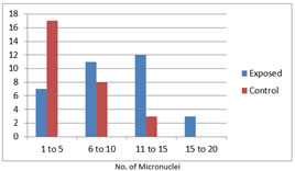
The mean and standard deviation (SD) of micronuclei in buccal cells in exposed was 8.28 and in controls was 1 and given in Table 2. There was a significant increase in the frequency of MN in exposed group when compared with the comparison group (p<0.001). The maximum number of exposed had a MN range between 11- 15 and the comparison group it was between 1- 5 (Figure 1).
Table 2: Mean and Standard Deviation of frequency of micronuclei in buccal cells
Figure 1: Frequency distribution of MN in Buccal cells of exposed and controls. Maximum number of exposed have a MN range between 11 - 15 and controls have 1-5. The mean and standard deviation (SD) of micronuclei in peripheral blood in exposed was 8.35 and in controls was 4.18 and given in Table 3. There was a significant increase in the frequency of MN in exposed group when compared with the comparison group (p<0. 5876). The maximum number of exposed had a MN range between 11- 15 and the comparison group it was between 1- 5 (Figure 2).
Table 3: Mean and Standard Deviation of frequency of micronuclei in peripheral blood
Figure 2: Frequency distribution of MN in Peripheral blood of exposed and controls. Maximum number of exposed have a MN range between 11 - 15 and controls have 1-5. Pearson’s correlation test was used to correlate between the duration of exposure and frequency of MN. There was a positive correlation between the years of FA exposure and the number of micronuclei in buccal cells and peripheral blood indicating that DNA damage due to FA was directly proportional to the duration of exposure (r=0.8, 0.9).
DISCUSSION Formaldehyde is a clear, colorless flammable strong-smelling chemical that is widely used in Anatomy laboratories mainly for embalming and also as a preservative. When the air level of formaldehyde exceeds 0.1ppm individual experiences adverse effects like burning sensation in eyes nose throat and skin irritation. Several studies have suggested that there is an increased risk of cancer particularly leukemia on long term exposure to formaldehyde than on general population. Micronuclei are the extranuclear bodies which are the chromosome fragments or the chromosomes that lag behind at the anaphase of the dividing cells and not incorporated into the nucleus of the dividing cells. It is commonly used to identify the population at risk after getting exposure to the carcinogens. Micronuclei can be studied in cultured lymphocytes and can be scored in binucleated cells in dividing eukaryotic cells.7 Various indicators are used to detect the cytogenetic alterations caused due to FA exposure. Among them Cytokinesis Blocked Micronucleus Assay (CBMA) is a sensitive and well-established tool for measuring the DNA damage because of its simplicity and rapidity.4 In the present study DNA damage was assessed in peripheral lymphocytes and in buccal cells of Formalin exposed workers in different laboratories. In the present study the mean and standard deviation of micronuclei in the peripheral blood was 8.35 and 5.03 respectively with a p value of 0.58. It was similar to the study conducted by D souza et.al where the mean and SD was 9.5+3.23 with a significant p value.1 In a study by Costa et.al on the genotoxic effects of formaldehyde by using comet assay and the micronuclei in the peripheral blood the mean and SD was 6.19+0.62 where they also studied the level of exposure by using time weighted average which exceeded the national and international levels of 0.3 ppm.3 In a study by Viegas et.al the micronucleus in the peripheral blood was 3.7+3.8 among those exposed in anatomy and pathology laboratories and among the factory workers it was 1.76+2.07 thereby stressing the fact that this information is important for risk assessment process and may be used to assess health risks for exposed workers8 Buccal epithelial cells remain the preferred site for the diagnosis of genotoxic effects of the carcinogenic agent. The basal layer of the epithelial cells undergoes damage where the cells undergo mitosis. Because of rapid turnover of the epithelial tissue the cells exfoliate to the surface. So, detection of micronucleus in the buccal epithelial cells has advantages like limited cost, can be counted easily person time required is less and precision obtained from scoring large number of cells makes this noninvasive method very popular.4 In the present study the mean and SD of micronucleus in the buccal cells were 8.29 and 4.69 respectively with a p value of 0.0001 which was statistically significant. It was similar to the study conducted by D’souza et.al where the mean and SD of the micronucleus were 9.5+3.23 with a P value < 0. 001.(1)In a study by Viegas et.al who studied the genotoxic effect of formaldehyde exposure among the laboratory as well as factory workers exposed to formaldehyde the frequency of micronucleus was high among the exposed group with a P value < 0.001 which stress the point that risk assessment of the health for the exposed group should be done frequently.8 In a study by Lorenzoni et.al who investigated the mutagenicity of FA in buccal cells among the anatomy students before exposure to formalin and 1 to 3 months after exposure which showed that there was a steady increase in the frequency of micronucleus before and after exposure.4 In the present study the duration of exposure ranged between 1-30yrs with the mean duration of 9 yrs.There was a positive correlation between the years of FA exposure and the number of micronuclei in buccal cells and peripheral blood indicating that DNA damage due to FA was directly proportional to the duration of exposure (r=0.8, 0.9). It was similar to D’Souza et al. who showed a positive correlation between the years of exposure and frequency of micronuclei in lymphocytes (r=0.5, p = 0.02).1
CONCLUSION The present study was done to assess the DNA damage in people who were exposed to FA and a control group not exposed to FA by buccal cell and peripheral blood Micronucleus Assay. There was a significant increase in the MN in people exposed to FA which was directly proportional to the duration of exposure.
REFERENCES
Policy for Articles with Open Access
|
|
||||||||||||||||||||||||||||||||||||||||||||||||||||||||||||||||||||||||

 This work is licensed under a Creative Commons Attribution-NonCommercial 4.0 International License.