 Home
Home|
Table of Content Volume 4 Issue 2 -November 2017
Variation of bilateral superficial palmar arch – A case report with its clinical significance
Sonal Govindwar1, Swapna Chavan2*
1Assistant Professor, Nanded Rural Dental College & Research Center, Nanded, Maharashtra, INDIA. 2Assistant Professor, Department of Anatomy, HBT Medical College and Coopar Hospital, Vile Parle, West Mumbai, Maharashtra, INDIA. Email: sonal.govindwar@gmail.com, swapna.chavan2012@gmail.com
Abstract The anastomoses between radial and ulnar arteries in the palm play a significant role in diseases of the palm through collateral circulation. During routine dissection of the upper limb of a 45-year-old male cadaver, we observed the bilateral incomplete superficial palmar arch (SPA), where both the superficial branches of radial and ulnar artery were present but completely fails to join with each other to form a complete SPA. In right hand, the superficial palmar branch of the radial artery entered the hand above the thenar muscles and provided palmar digital branches to the radial side of the index finger and the ulnar side of the thumb, without any contribution to the SPA. The superficial branch of the ulnar artery gave origin to three common palmar digital arteries to supply the contiguous sides of the index, middle, ring and little fingers. In left hand, both the arteries did not give any branches and ends in the straight course without anastomosis. Key Words: Radial artery, Superficial palmar arch, Ulnar artery.
INTRODUCTION The anastomoses between radial and ulnar arteries in the palm play a significant role in diseases of the palm through collateral circulation. During routine dissection of the MBBS students we have seen that, the both hand of a 45 year-old male cadaver, showed variation in the formation of the superficial palmar arch (SPA). [The superficial arteries of the hand formed several diversified patterns that permitted into well-defined categories. About two-third of the SPA is formed by the ulnar artery alone; a further third is completed by the superficial palmar branch of the radial artery and a third either by the arteria radialis indicis or by the princeps pollicis or by the median artery. A classic type of SPA in which the superficial branch of the radial artery joins the superficial branch of the ulnar artery is found only in 34.5% of the cases. There are many reports regarding formation of SPA. In a study by Coleman et al.1, the complete arch was found in 78.5% of the cases and incomplete arch in the remaining 21.5%, and this formed a major underlying factor in the aetiology of digital ischaemia. Ikeda et al2. conducted stereoscopic arteriography of 220 cadaver hands and reported complete SPA in 96.4% of the cases, and only 3.6% had an incomplete arch. Gellman et al. showed a complete SPA in 84.4% and Al Turk and Metcalf reported complete SPA in 84% of the cases.
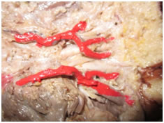
CASE REPORT In the present case, we observed following types of variation in superficial palmar arch. Figure 1: Showing the incomplete right superficial palmar arch Figure no. 1 showing the variations in the formation of superficial palmar arch on right side, where both the branches i.e. superficial palmar branch of the radial and ulnar artery were present but, fail to anastomose with each other. The superficial palmar branch of the radial artery entered the hand through the thenar muscles at the flexor polices brevis and passed superficial to the thenar muscles and provided palmar digital branches to the radial side of the index finger and the ulnar side of the thumbSuperficial branch of ulnar artery was giving three branches and superficial branch of radial artery was giving off two branches. Figure 2: Showing the incomplete of left superficial palmar arch
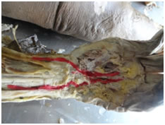
Figure No. 2 showing the incomplete superficial palmar arch on left side. Here, the superficial branches of radial and ulnar were present without any branching, but they did not unite with each other, thus there was absence of superficial palmar arch. Deep palmar arch in both the hands was normal.
DISCUSSION Figure no. 1 and 2, was showing the cases where the both the superficial palmar branches of radial and ulnar artery were present, without anastomosing with each other. This type of variation was classified as a type A of group II, as per the Coleman and Anson (1961)1 classification. This type of variation was observed by patnaik, V.V.VG, et al (2002)3, in 3.2% cases, suleyman murat tagil et al (2007)4, in 3.2% cases, sanjay kumar Sharma et al (2013)5, in 10% cases, where as Challa. Ratna Prabha et al (2014)6 presented a case report where this type of arch was observed. In present study such type of arch was observed in 13.4% cases. The explanations for the arterial variations are based on classical outline of arterial development. Arey7 is of the view that the anomalies of blood vessels may be due to the choice of the unusual paths in the primitive vascular plexuses, the persistence of vessels normally obliterated, incomplete development or absorption of the parts usually distinct.
CONCLUSION The existence of both common and rare anatomic variations in the formation of superficial palmar arch as well as the absence of collateral circulation between ulnar and radial arteries necessitates the proper knowledge of vasculature of the hand in order to avoid or minimize the risk of complications during vascular surgeries or reconstructive surgeries in the hand.
REFRENCES
|
|
 This work is licensed under a Creative Commons Attribution-NonCommercial 4.0 International License.