 Home
Home|
Table of Content Volume 7 Issue 1 - July 2018
A study of cuboidal articular surface of calcaneus and its clinical significance
Eluru Ravitheja1, Dasari Sudhakar Babu2*, Siraz M Ausavi3
1Associate professor, 3Professor and HOD, Great Eastern Medical School, Ragolu Village, Srikakulam district, Andhra Pradesh- 532484 INDIA. 2Associate Professor, Department of Anatomy, Government Medical College, Nizamabad, Telangana. 503001 INDIA.
Abstract Aim: Calcaneus is the largest and most frequently fractured tarsal bone. The cuboidal articular surface is concavo-convex which fits on to the reciprocal surface of cuboid. The aim of study is to observe the different shapes of cuboidal articular surface of calcaneus and their importance in clinical aspects of ankle instability Materials and Methods: 60 dry human calcanei (25 right, 35 left) of unknown sex collected from the bone bank of Anatomy department, Great Eastern Medical School, in Srikakulam district of Andhra pradesh were used for the study. The shape of the cuboidal articular surface of various calcaneii were studied and results tabulated. Results: Wedge (42%) and Vertically oval (23%) shaped articular surfaces were seen in more number of calcaneii and the other shapes include Triangular, Rectangular, Circular, Irregular and Comma shaped surfaces. Conclusion: The knowledge of shape of the cuboidal articular surface of the calcaneus is useful for radiologists and orthopaedic surgeons in permorfing arthoscopic peroneal tendon reefing for correcting chronic lateral ankle instability and in triple arthodesis for club foot. Key Words: Cuboidal articular surface, ankle instability, calcaneus.
The calcaneus is the largest of the tarsal bones and projects posterior to the tibia and fibula as a short lever for muscles of the calf attached to its posterior surface. It is irregularly cuboidal, its long axis being inclined distally upwards and laterally1. It takes part in transmission of the body weight and helps in forming three joints namely, talo-caloaneal, calcaneo–cuboidal, talocalcaneonavicular joints2. The anterior surface is smallest and is obliquely set to articulate with concavo-convex articular facet of cuboid. The lateral surface is flat it is proximally deeper and palpable on the lateral aspect of the fibular trochlea (peroneal tubercle) which is exceedingly variable in size and palpable 2cm distal to the lateral malleolus when well developed. It bears an oblique groove for peroneus longus and a shallow proximal groove for attachment of the calcaneofibular part of the lateral ligament 1. Primates have significantly elongated calcaneus in comparision to many non-primates 3. The cuboidal articular surface on the calcaneus is one of the most distinctive feature of the human foot in relation to the long axis of the calcaneus. The superior margin of human cuboid facet projects further anteriorly than does the inferior margin. There is generally a relatively deep concavity on the medial aspect of facet which articulates with the beak like projection on the articular surface of cuboid. From this medial convexity, the calcaneal articular facet flares out laterally in a moderately convex surface that has been termed the anterolateral articular process of the calcaneus. The complex shape of this joint surface allows the distinctive lateral swing of the human calcaneocuboid joint in to a close packed position during the stance phase of the bipedal locomotor cycle and allows the midtarsal region of the foot function as a rigid lever4.
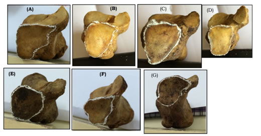
MATERIALS AND METHODS 60 dry human calcaneii (25right, 35 left) of unknown sex were collected from the bone bank of Anatomy department, Great Eastern Medical School, in Srikakulam district of Andhra Pradesh. All the bones were studied for the following variables, presence of notches on the lateral margin of cuboidal articular suface and their number, distance of the notch from the lowest point of the facet, total height of fac et al long the lateral margin of the facet, the type of peroneal tubercle and its relation to the presence of notch, the shape of the cuboidal articular facet. Measurements were taken with a sliding callipers calibrated to 0.1mm Different kinds of cuboidal articular surfaces are shown in fig.1
Figure 1: A) Wedge -42%, B) Vertically oval-23%, C) Triangular-13% D) Rectangular-3%, E) Circular-10%, F) Irregular-7%, G) Comma shaped-2%
DISCUSSION Ankle sprains are one of the most common lower extremity injuries although most people recover without significant long-term complications. Chronic ankle instability does develop in about 20% of patients5,6,7. Affected individuals usually complain of recurrent ankle sprains, difficulty with ambulation on uneven ground and pain with activity. Peroneal tendon injuries are at increased risk of chronic ankle instability8,9. While performing an arthroscopic repair it is important to have a good understanding of the proximity of surrounding structures. There is a paucity of anatomic studies defining safe zones and structures at risk for arthoscopic repair10. Christopher F. Hyer et al in 2005 classified peroneal tubercle structurally as flat prominent in 29% concave in 27.2% and tunnel in – 1.0%11. Agarwal AK, et al studied the variation in structure of human calcanei of people of Agra and Lucknow12. Jyothi KC, shailaja shetty from Karnataka classified human calcaneii based on the cuboidal articular facet into wedge, vertically oval, horizontally oval, and irregular13. Study of relationship between the type of peroneal tubercle and notch on the lateral margin of the cuboidal articular surface of calcaneus was never attempted in the past. This data may help orthopedic surgeons in ankle arthoscopic procedures to be done on patients of Andhra pradesh.
CONCLUSION The knowledge of the relation between the notch on the lateral margin of the cuboidal articular surface of calcaneus is useful for radiologists and orthopedic surgeons in tendonitis of peroneus longus and peroneus bervis, in repositioning the dislocated peroneal tendons and also in treating the chronic lateral ankle instability by ankle arthoscopy. The knowledge of shape of articular surface is useful for doing corrective osteotomy and triple arthodesis for club foot.
REFERENCES
|
|
 This work is licensed under a Creative Commons Attribution-NonCommercial 4.0 International License.