 Home
Home|
Table of Content Volume 7 Issue 3 - September 2018
A study on morphometric analysis of foramen ovale among south Indian population
Durai Pandian K1, K V K Sundaravadhanam2*, T L Anbumani3
1Associate Professor, 2Assistant Professor, 3Professor, Department of Anatomy, Karpaga Vinayaga Institute of Medical Sciences, Chinna Kolampakkam, Maduranthagam, Kanchipuram, District, Tamil Nadu, INDIA. Email: drkvksundar@gmail.com
Abstract Background: Greater wing of sphenoid has many foramens, of which foramen ovale is of great importance as mandibular nerve passes through it, any alterations from the normal foramen leads to vascular compromise and nerve compression. Materials and Methods: 40 dry adult human skulls were chosen during routine demonstration to undergraduate medical students. The Foramen ovale of the entire chosen specimens was evaluated in terms of size, shape using Vernier caliper from the base of the skull. Results: The mean length of foramen ovale on right side is (6.93mm ±0.85mm) and (6.88mm ±0.87mm) on left side. The mean breadth of foramen ovale on right side is (3.99mm ±0.59mm) and (3.85mm ±0.56mm) on left side. Oval shaped foramen ovale was about 65% on right side and 75% on left side, followed by almond shape was about 17% on right side and 12% on left side and circular shape was about 17% on right side and 12% on left side. Conclusion: The study has made it evident that the different shapes of foramen might be a reason for nerve compression, further studies with larger sample size and comparing them with real clinical patients will throw light on other morphological variations which would help clinicians for diagnosis and treatment of Neuralgia. Key Words: Foramen Ovale, TrigeminalNeuralgia, Skull, Sphenoid bone.
Foramen ovale is an essential foramen present on infratemperol surface of greaterwing of sphenoid. It transmits various neurovascular structures which include mandibular nerve, lesser petrosal nerve, accessory middle meningeal vessels and emissary vein1. Foramen ovale has great surgical and clinical significance in nerve compression causing trigeminal neuralgia and to perform percutaneous procedure for cavernous sinus by clinicians. Many eminent researchers have studied about the morphometrics of foramen ovale based on shape and length; the knowledge of anatomy of foramen ovale is not only helpful for anatomist but also useful for radiologist, orofacial-maxillary surgeons and neurosurgeons. The present study was carried out to identify various morphometric analysis of foramen ovale to identify the variations in the morphology so as to aide in the diagnosis of compression leading to neuralgia.
MATERIALS AND METHODS This study was performed in 40 dry adult human skulls during routine demonstration for undergraduate students in the department of Anatomy, Karpaga Vinayaga Institute of Medical Sciences, Madhuranthagam, Kanchipuram District, Tamil Nadu, India. Parameters like shape and size were studied using Vernier calipers from the base of the skull. The shape of foramen ovale on both sides was measured. Maximum length and breadth was calculated using Vernier caliper. The results were analyzed statistically using SPSS version 20.The study was cleared by internal ethical committee.
RESULT All the skulls showed bilateral presence of foramen ovale. The frequency of oval shaped foramen was maximum about 65% on the right side and 75% on the left side. The frequency of almond shape was 17.5% on right side and 12.5% on left side. The frequency of circular shape was 17.5% and 12.5%.The mean length and breadth of foramen ovale on right side was 6.93mm and 3.99mm.The mean length and breadth of foramen ovale on left side was 6.88mm and 3.85mm.
Table 1: Shape of Foramen Ovale on the right side
Table 2: Shape of Foramen Ovale on the left side
Figure 1: Bar Diagram of Shape Vs Side
Table 3: Chi square tests to find the association b/w right and left side in shapes at 5%α
There is no association b/w right and left side of shapes, Breath right vs. left = 33.9%, p = 0.03**, Breath vs. Shape = - 36 % with p = 0.001*, *Significant by Spearman Rank correlation. There is significant relationship between the two variables, ** Significant by Kendal’s correlation. There is significant relationship between the left and right side. Table 4: Paired t test to find the diff b/w two related variable at 5%α
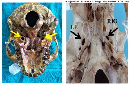
There is no significant difference b/w right and left side in length. There is no significant difference b/w right and left side in breath. Figure 1 Figure 2 Figure 1: Showing circular FO on both sides; Figure 2: Showing almond shaped FO in Left and oval shaped FO on Right DISCUSSION Complete knowledge of development of foramen ovale is essential to identify the variations and abnormal foramens. The greater wing of sphenoid is embryologically complex structure developed by endochrondral ossification by a piece of cartilaginous bar called Allispenoid which encloses the mandibular nerve and other vasculature to form greater wing of sphenoid. A complete ring shaped foramen ovale is seen in 7th month and 3rd year2. Many authors have demonstrated about the variations of shape of foramen ovale. Maximum percentage of oval shaped of foramen ovale was demonstrated by many authors, thus correlating with our study and in greys anatomy as the shape the foramen ovale is oval1. Daimi et al have showed 46%of D shaped foramen ovale8. The comparative study of various authors is tabulated in table 5.In the present study the shape of foramen ovale was 65% oval on right side and 75% on left side followed by almond shaped (17.5% in right side and 12.5% in left side). The percentage of circular shape was 17.5% in right side and 12.5% on left side. Thus any alteration of shape leads to nerve compression.
Table 5: Comparative study of shape of foramen ovale in previous study
Study among Nigerian population showed a mean length ranging from 9.5mmto 5mm12. Study among Mangalore population showed a mean length 7.6mm to 7.01mm3. Landl et al studied among new york population using Fluoroscopic assisted laser showed a mean length of 6.9mm on right side and 6.8mm on left side13. A study among Kenyan population showed a higher values of mean length and breadth. Comparative study of morphometric analysis has been shown in table 6.Morphometric analysis of foramen ovale in the present study showed mean length (6.93 ±0.85) on right side and (6.88 ±0.87) on left side and mean Breadth (3.99 ±0.59) on right side and (3.85 ±0.56) on left side. There was on statical significance between the right and left side based on length and breadth. Table 6: Comparative study of morphometric analysis of foramen ovale by various authors
Investigative procedures like fine needle aspirationcytology through transfacial approach, Percutaneous trigeminal rhizotomyare performed through Foramen ovale. The normal anatomy of foramen ovale has a great clinical and surgical significance.
CONCLUSION Foramen ovale is inconsistant in shape having a maximum percentage of oval shape, that is, 65% on right and 75% on left. The mean length was around 6.9mm on right side and 6.8mm on left side. The mean breadth was around 3.9mm on right side and 3.8mm on left side The study has made it evident that the different morphology of the foramens might be a reason for nerve compression, further studies with larger sample size and comparing them with real clinical patients may throw light on other morphological variations which would help clinicians for diagnosis and treatment of Neuralgia
REFERENCES
|
|
||||||||||||||||||||||||||||||||||||||||||||||||||||||||||||||||||||||||||||||||||||||||||||||||||||||||||||||||||||||||||||||||||||||||||||||||||||||||||||||||||||||||||||||||||||||||||||||||||||||||||||||||||||||||||||||||||||||||||||||
 This work is licensed under a Creative Commons Attribution-NonCommercial 4.0 International License.