 Home
Home|
Table of Content Volume 8 Issue 1 - October 2018
Study of variations in the draining pattern of pulmonary veins in left atrium
Bhingardeo A V1*, Bhoir M2, Mundada K V3
1Registrar, 2Professor, 3Second MBBS student, Department of Anatomy, HBTMCand Dr. R. N. Cooper Municipal General Hospital Mumbai, Maharashtra, INDIA. Email: dr.alkabhingardeo@gmail.com
Abstract Background: Pulmonary veins carry blood from lungs to the left atrium. Normally four pulmonary veins are present (2 right and 2 left). Variations in the number of pulmonary veins act as electrical focus for triggering arrhythmia. In the present study we evaluated draining pattern of pulmonary veins in 33 formalin fixed hearts Aims and objective: To study variation in draining pattern of pulmonary veins in 33 cadaveric hearts Materials and method: We studied the draining pattern of pulmonary veins into left atrium in 33 formalin fixed hearts. Pulmonary venous openings in left atrium were observed and categorized as right superior, right inferior, left superior and left inferior. When more than four pulmonary veins were found then the number of pulmonary veins and the manner of their opening into the left atrium was noted. Results: We found variations in 13 hearts out of total 33 hearts. Variations were more common on right side (39.39%) than on left side (9.09%). Maximum number of pulmonary veins reported on right side were 4(12.12%) whereas on left side were 3 (3%). Most common variation on right side was the presence of 3 pulmonary veins with 3 ostia which is found in 18.2% of cases while on left side the most common variation was the presence of three pulmonary veins with 2 ostia which is found in 3% of cases. Conclusion: Variation in draining pattern of pulmonary veins is not rare. Knowledge of pulmonary venous anatomy will be helpful for radiologist and surgeons for finding exact ectopic focus causing arrhythmia in radiofrequency ablation procedure for arrhythmia. Key Word: pulmonary veins, left atrium, arrhythmia
INTRODUCTION Pulmonary veins are one of the major structures of the circulation1. Approximately 70% of the general population has four pulmonary veins which are responsible for bringing the oxygenated blood from the lungs to left atrium2,3,4 They develop as an outgrowth of the dorsal atrial wall just lateral to the septum primum in the sinoatrial region.5,6, 7 There are two superior and two inferior pulmonary veins. Normally these veins open in the left atrium at their independent ostia. Anatomical variations in the number and branching pattern of the pulmonary veins result from the under incorporation or over incorporation of the common pulmonary vein.5 The variation in the pulmonary venous anatomy results in ectopic beats. These veins are important source of ectopic electrical activity frequently initiating paroxysms of atrial fibrillation. 8Radiofrequency ablation of such arrhythmogenic foci is carried out as a part of treatment.9 For such invasive procedures the detailed knowledge of pulmonary venous anatomy and relationship between pulmonary veins and left atrium is necessary. The objective of present study was to evaluate the variations in the draining pattern of pulmonary veins into the left atrium.
MATRIALS AND METHOD The present study was carried out on 33 formalin fixed hearts obtained from the Department of Anatomy of a tertiary health care center, Mumbai. Damaged hearts, hearts with grafts or previous surgery were excluded from the study. The left atrium was examined for the ostia of pulmonary veins opening into it. The pulmonary veins were grouped as superior (right and left) and inferior (right and left). The number of right superior, right inferior, left superior and left inferior pulmonary veins were noted. When more than four pulmonary veins were found then the number of pulmonary veins and the manner of their opening into left atrium was noted.
OBSERVATION AND RESULT Table 1: Number of hearts showing variation in the number of pulmonary veins
Table 2: Variations in Right pulmonary veins
Table 3: Variations in Left pulmonary veins
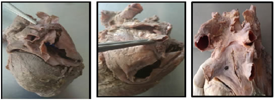
Figure 1 Figure 2 Figure 3 Figure 1: Three superiorright pulmonary veins; Figure 2: Four inferior right pulmonary veins; Figure 3: Two right superior and two right inferior pulmonary veins. DISCUSSION Accessory pulmonary veins occur by over incorporation of the pulmonary veins beyond their first division. Such variations in draining pattern of pulmonary veins act as ectopic focus causing cardiac arrhythmia12 We found variations in 13 hearts out of 33.Variations were more common on right side (39.39%) than on left side (9.09%). This is in agreement with studies by other authors.2, 3 Prasanna et al13 found variations in right pulmonary veins in 28% of cases while in left pulmonary veins in only 6% of cases while Kaur et al8 found 26% of cases who showed variations on right side while 23% of cases showing variations on left side. Most common variation in right pulmonary vein was the presence of 3 veins with 3 ostia. It was found in 18.2% of cases. Marom et al10 found the same variation in 24% of cases while Lovesh shukla11 et al found it in 6.9% of cases. Maximum number of veins on right side was 4 which is found in four cases in the present study. Prasanna et al13 and other authors reported the maximum number of veins up to five. The draining pattern of left pulmonary veins in maximum cases (96.96%) were normal with 2 pulmonary veins with 2 ostia. This result matches with findings of Marom et al10 (86%) and Prasanna et al13 (79.3%). The most common variation found on left side was the presence of 3 pulmonary veins with 2 ostia which is found in 3% of cases. Lovesh shukla11 found the same variation in 3.4% of cases. Out of total 13 variations, three hearts showed drainage of accessory veins by forming common stem while in remaining ten cases, accessory veins were found to have independent ostia. Variation in number of pulmonary veins leads to Cardiac arrhythmia which is the most common cause of cardiac morbidity and mortality8 Therefore knowledge of pulmonary venous drainage pattern is necessary. Radiofrequency ablation procedure is carried out for such ectopic foci resulting from variant pulmonary veins.9 The information regarding pulmonary venous drainage guides interventionist to reach the specific location of ectopic tissue of atrial activity and reduce the complications of missing ectopic foci in radiofrequency ablation procedures.8
CONCLUSION The findings of present study confirmed that there are variations in the number and draining pattern of pulmonary veins. Knowledge of such variations is necessary for radiologist and thoracic surgeons before doing any procedure involving pulmonary veins like radiofrequency ablation procedure for cardiac arrhythmia, pulmonary lobectomy, cardiac valve replacements to avoid post post procedural complications.11,14
REFERENCES
|
|
||||||||||||||||||||||||||||||||||||||||||||||||||||||||||||||||||||||||||||||||||||||||||
 This work is licensed under a Creative Commons Attribution-NonCommercial 4.0 International License.