 Home
Home|
Table of Content Volume 8 Issue 2 - November 2018
Incidence of accessory head of flexor pollicis longus (Gantzer’s) muscle
Raja Sekhar Katikireddi1, Raju Sugavasi2*
1Associate Professor, Department of Anatomy, Bhaskar Medical college, Yenkapally (V), Moinabad mandal , Ranga Reddy District, Telangana. 2Assistant professor, Department of Anatomy, Fathima Institute Of Medical Sciences (FIMS), Kadapa , Kadapa District, Andhra Pradesh. Email: anatraju@yahoo.co.in
Abstract Background: The additional head of the flexor pollicis longus muscle is also known as the Gantzer’s muscle. The present study was designed to evaluate the Accessory Head of the Flexor Pollicis Longus muscle and its incidence in south Indian population. Materials and Methods: 40 number adult upper limbs were used to study the additional heads of Flexor Pollicis Longus muscle. Results: Present study found 4 cases of additional heads of Flexor Pollicis Longus muscle in unilaterally out of 40 limbs. The incidence of the Gantzer muscle was recorded as 10% and Double Gantzer muscle bellies were also found in one limb out of 40 upper limbs and incidence was 2.5%. Conclusion: An accessory muscle bellies are the reasons for neuropathies of different types. Key Word: Flexor pollicis longus, Additional Head of Flexor pollicis longus), Gantzers muscles, Anterior interosseous nerve, Median nerve
INTRODUCTION Flexor pollicis longus (FPL) muscle is one of the deep flexors of the forearm. It takes its origin from the grooved anterior surface of the radius and from the adjacent interosseus membrane and gets inserted onto the base of the distal phalanx of the thumb1.The accessory head of the flexor pollicis longus muscle (AHFPL) is called as Gantzer’s muscle, this muscle arises as small belly from forearm flexors and is inserted either into Flexor Pollicis Longus or Flexor Digitorum Profundus2. Bilateral occurrence of Gantzer’s muscle is most common than unilateral presence3,4,5. The incidence of the AHFPL belly is probably from the development of the common flexor mass in the embryo and incomplete differentiation of it can creates the AHFPL6. Paralysis of the forearm due to compression of AIN in the forearm is also called as the Kiloh-Nevin syndrome7, 8.
MATERIALS AND METHODS Present study was conducted at Department of Anatomy, Bhaskar Medical College, Telangana. Total of 40 upper limbs of 20 adult cadavers were used for this study. Anterior compartment of forearms were dissected either side of cadavers by exposing the superficial and deep compartments of forearms. Muscles of forearm examined by their attachments, relations and nerve supply.
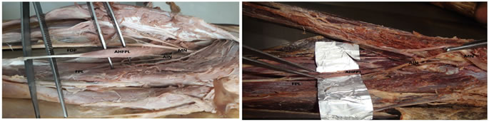
RESULTS Additional heads of flexor pollicis longus (AHFPL) muscle bellies were found in left side of 2 upper limbs (Figure. 1 and 2) and one on right side (Figure. 3) of upper limb unilaterally. Double bellies of AHFPL (Figure. 4) were noticed in left side unilaterally in one limb. All the AHFPL muscle heads were seen below the flexor digitorum superficialis and inserted to the flexor pollicis longus muscle. The Median nerve (MN) is passes anterior and lateral to the Gantzers muscles and Part of anterior interoseseous nerve (AIN) is observed posterior to the muscle and in all cases additional bellies were supplied by anterior interosseous nerve. Incidence of Additional muscle bellies of present study was noted as10% (4 out of 40) and Double Gantzer muscle incidence was 2.5% (1 out of 40). 4(10%) out of the 40 dissected forearms in the present study. Double Gantzer muscle bellies were found in 1 out of 40 limbs and incidence was 2.5%.
Figure 1 Figure 2 Figure 3 Figure 4
Figure 1: Left Forearm shows, AHFPL: Additional Head Of Flexor Pollicis Longus, FPL: Flexor Pollicis Longus, FDP: Flexor Digitorum Profundus, MN: Median Nerve, AIN: Anterior Interosseus Nerve.; Figure 2: Left Forearm shows, AHFPL: Additional Head Of Flexor Pollicis Longus, FPL: Flexor Pollicis Longus, MN: Median Nerve, AIN: Anterior Interosseus Nerve; Figure 3: Right Forearm shows, AHFPL: Additional Head Of Flexor Pollicis Longus, FPL: Flexor Pollicis Longus, FDP: Flexor Digitorum Profundus, MN: Median Nerve, AIN: Anterior Interosseus Nerve. Figure 4: Left Forearm shows, AHFPL: Additional Head Of Flexor Pollicis Longus, FPL: Flexor Pollicis Longus, FDP: Flexor Digitorum Profundus, MN: Median Nerve, AIN: Anterior Interosseus Nerve. The additional muscle belly was found in the present study was the accessory head of flexor pollicis longus (FPL). Incidence of such additional head of FPL in Indian population was reported previously by various authors; Malhotra et al., (1982)9 incidence was 54.2%. Hemmady et al., (1993)10 observed 36 cases out of54 limbs and incidence was (66.67%). Pai et al., (2008)11noticed 58 in 126 limbs (46.03%). Sembian et al., (2012)12 incidence was (0.50%). Tamang et al., (2013)13found 15 out of60 cases (25.0%). Gunnal et al., (2013)14observed 92 out of 180 limbs and the incidence was (51.11%). V Bangarayya et al., (2018)15 noticed 12 Out of 30 upper limbs and prevalence was (40%). The incidence of the Gantzer muscle was recorded in 4(10%) out of the 40 dissected forearms in the present study. Double Gantzer muscle bellies were found in 1 out of 40 limbs and incidence was 2.5%. Compression of the anterior interosseous nerve (AIN) by this type of additional bellies could appear and give rise to anterior interosseous syndrome. AIN compression is seen to be more possible in literature than Median Nerve compression (Caetano et al., 2015)16.When entire anterior interosseous nerve passes underneath the Additional belly that causes weakness in the FPL, in the FDP of the index and the middle fingers, and in the pronator quadratus muscles. Presence of AHFPL can compressed the AIN that causing an isolated paralysis of the FPL with a characteristic pinch movement disability17.
CONCLUSION The present study found the incidence of occurring accessory head of flexor pollicis longus in south Indian population and with relation to anterior interosseous nerve (AIN). This study may beneficial for the orthopaedic surgeons while they perform de compressive fasciotomies for the forearm compartment syndrome.
REFERENCES
|
|
 This work is licensed under a Creative Commons Attribution-NonCommercial 4.0 International License.