 Home
Home
Official Journals By StatPerson Publication
|
Table of Content - Volume 12 Issue 3 - December 2019
Khushboo Rastogi1*, Himanshu Upadhyay2, Yukti Pawar3
1Associate Professor, 2Undergoing Post Graduation in (Periodontology and Implantology) Department of Dentistry, Saraswathi Institute of Medical Sciences, Flat No. 04, Ground Floor Staff Quarters, SIMS Anwarpur Hapur- 245101 Email: dr.khushboorastogi@gmail.com
Abstract Background: The purpose of this study was to evaluate the effectiveness of PRF in the healing outcome of mandibular mesioangular impacted third molar tooth, to assess post-operative pain, swelling and trismus, pocket depth on the distal aspect of the second molar, the degree of bone regeneration at the extraction site after six months and also to assess soft tissue healing. Methods: The study was designed as A Randomized controlled clinical interdepartmental study. A total of 40 patients who reported to the Out-Patient Department, fulfilling the inclusion criteria and who voluntarily consented to participate in this study were selected. Two study groups were formed that included a Group A (Control Group) that constituted of surgical removal of impacted mandibular third molar followed by flap approximation and a Group B (Test Group) that constituted of surgical removal of the impacted mandibular third molar, from which 10 ml venous blood was drawn and centrifuged at 3000 rpm for 10 minutes to prepare the Platelet Rich Fibrin, and then be placed into the extraction socket, followed by flap approximation. Patients were evaluated and compared preoperatively, immediately post operatively, at the end of one week, one month, three months, and 6 months. The outcome variables assessed were edema, trismus, pain, pocket-depth, bone density and soft tissue healing. A statistically significant difference was observed in the duration of surgery between the case and the control group in the present study. It could attribute to the fact that there was consumption of time in preparation of PRF during the surgical procedure. In the present study, interincisal distance was recorded in all patients preoperatively, on the second postoperative day, seventh postoperative day and at 1, 3 and 6 months postoperatively to assess restriction in mouth opening in the case and control group. A statistically significant difference was observed on the second and seventh postoperative day, and at 1 month follows up. There was no significant difference in interincisal distance at 3 and 6 months follow up. The present study also revealed that PRF significantly recorded less pain, swelling, increased bone density and fast soft tissue healing for the second and seventh postoperative days, whereas no differences were observed for the first, third and sixth postoperative months when compared with case and control group. Conclusion: The finding of present study concluded that PRF application after impacted mandibular third molar surgery is a good biologic material to avoid post-operative sequalae like pain, trismus, swelling. We found definite clinical advantages with respect to the healing of soft tissues. As far as bone was concerned, there was an increase in quantity in the experimental group throughout the study period. And regarding pain and trismus we found less trismus and less pain as compared to control group. Thus Platelet-rich fibrin offers a natural way of augmenting soft and hard tissues. Key Words: PRF, Third Molar, Mesioangular, Impaction, Pain. Trismus, Pocket Depth and Soft tissue healing
INTRODUCTION The third molar is the most widely discussed tooth in the dental literature.1 It generally erupts in the oral cavity between the ages of 17 - 25 years. Impaction may be defined as the failure of complete eruption of a tooth into its normal functional position within the normal time span.2 The mandibular third molar is the most commonly impacted tooth.3 Third molars get impacted due to inadequate room in the dental arch for their eruption.4 Indications for the removal of impacted lower third molars are numerous and include recurrent pericoronitis, caries of the third molar and/or adjacent second molar, periodontal causes, orthodontic and prosthetic concerns and associated pathologies as the most common causes.5,6 The quality of life experienced by patients following third molar surgery is increasingly becoming a health concern.7 Third molar surgeries are associated with unpleasant experience by the patients, referred to as postoperative morbidity, which could be divided into immediate postoperative tissue reactions and complications.8,9 PRF in various surgical procedures such as management of intraosseous defects, grade II furcation, sinus floor augmentation during implant placement, with coronally displaced flaps in multiple gingival recession and in facial plastic surgery procedures has shown promising results. The existing literature mainly deals with the use of PRP in extraction sockets as well as in other areas with non-placement in control sites. However, there is limited information available concerning the healing of extraction sockets using PRF. The goal of this study was to evaluate the effectiveness of PRF in the healing outcome of mandibular mesioangular impacted third molar tooth.
METHODS A total of 40 patients who reported to the Out-Patient Department, fulfilling the inclusion criteria and who voluntarily consented to participate in this study were selected, which were randomly divided into two study groups of 20 each; Group A (Control group): After surgical removal of impacted mandibular third molar, only flap approximation was done. Group B (test group): After surgical removal of the impacted mandibular third molar, 10 ml venous blood was drawn and centrifuged at 3000 rpm for 10 minutes to prepare the Platelet Rich Fibrin, which was then placed into the extraction socket, followed by flap approximation. Patients were evaluated and compared preoperatively, immediately post operatively, at the end of one week, one month, three months, and 6 months. Preoperative Assessment A complete medical history was elicited and an oral examination was performed, including a radiographic evaluation, to confirm the need for mesioangularly impacted third molar removal. All patients were given full information regarding the purpose of the study and effects of the drugs used and written consent was obtained for participation in the study. Impacted mandibular mesioangular third molar indicated for surgical extraction were assessed for its angulation, position and depth by means of an intraoral periapical radiograph and/or panoramic radiograph and its Pederson’s difficulty score was calculated and 5-6 score were included in the study. Baseline data were recorded at the initial visit. Obtaining platelet rich fibrin (PRF) from the patient’s own blood: A day prior to surgery, the patients were screened through a complete hemogram analysis which included the platelet count, hemoglobin concentration, bleeding time, clotting and total and differential counts were done. Also screening for Australian antigen and HIV infection was also done. Next day, at the time of surgery, blood was drawn by venipuncture from the antecubital fossa of the patient’s forearm using a 24-gauge needle and collected into two blood collected tubes (vacutainers) having capacity of 5 ml each. Preparation of platelet rich fibrin(PRF) membrane The PRF was prepared following the protocol developed by Choukroun et al. (2001)4 before surgery, 10 ml of intravenous blood sample were taken in two test tubes which were immediately centrifuged at 3000 resolutions/min for 10 minutes. At the end of centrifugation, following three layers were seen: the top most layer of supernatant serum, the fibrin clot at the middle layer and the bottom layer contacting the red blood cells (RBC). This clot was removed from the tube and the attached red blood cells scraped off and discarded. The PRF clot was then placed on the grid in the PRF Box and covered with the compressor and lid. This produced an inexpensive autologous fibrin membrane in approximately 10 minutes. PROTOCOL FOR SURGERY: Surgery was performed by a single operator on all patients using a standard protocol. The patient’s face was scrubbed with betadine solution and then the patient was draped under sterile aseptic conditions. Anesthesia was achieved by a standard inferior alveolar nerve block and long buccal nerve block using a solution of 2% lignocaine hydrochloride and adrenaline bitartrate of 1:80000 ratios. Standard Ward’s incision was made in all the cases. Full thickness mucoperiosteal flap was elevated to expose sufficient bone on the buccal and distal aspect of the impacted molar. Bone was then removed around the tooth with a rotary round and straight fissure bur on a straight hand piece under continuous copious irrigation with sterile saline solution. This was done to create a gutter around the buccal and distal aspects of the tooth. Odontectomy was performed whenever necessary to facilitate tooth removal and the tooth was surgically removed. Wound toilet: After complete extraction of the tooth, the socket was thoroughly inspected, curetted for follicular tissue removal and any sharp bony edges were smoothened. Copious irrigation was done with sterile saline solution. The irregular margins of the wound were trimmed and the flap was closed with 3-0 black braided interrupted silk sutures. A small pressure pack was then applied to the site and Platelet rich fibrin was inserted in the group B patient prior to wound closure. Standard protocols for post extraction instructions were given to all patients. Additionally, all patients were asked to report on the 2nd and the 7th post-operative day, 1 month, 3 months, 6 month postoperatively for evaluation. ASSESSMENT AND FOLLOW-UP: Each subject was recalled for evaluation on 2nd and 7th day postoperatively: Measurements of edema: Edema on the operated side was evaluated by a 3-0 silk thread used to record the following distances described by Gabka and Matsumara. Using 5 reference points: tragus, soft tissue pogonion, lateral canthus of the eye, angle of mandible and outer corner of mouth, 3 measurements were made:
The preoperative sum of all these three measurements was considered as the baseline of that side.
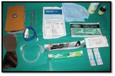
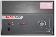
DATA ANALYSIS Data analysis was carried out using Statistical Package for Social Science (SPSS, V 11.5) package. Un-paired Student t-test was used to compare the mean of two groups(For statistical significance). In all the tests the “p” value of less than 0.05 was accepted as indicating statistical significance. PHOTOGRAPHS Figure 1 Figure 2 Figure 3 Figure 1: ARMAMENTARIUM FOR PREPARATION OF PRF; Figure 2: Centrifugal Machine; Figure 3: Surgical armamentarium
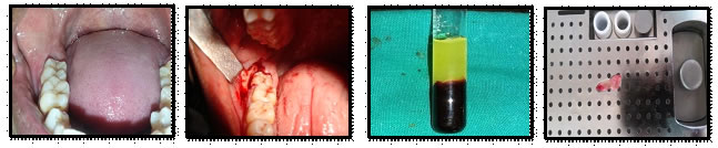
INTRAOPERATIVE PICTURES WITH PRP Figure: 04 Figure: 05 Figure: 06 Figure:07 Figure 8 Figure 9 Figure 10
FIGURE 04: INTRA ORAL POSITION OF 3rd MOLAR, FIGURE 05: BONE GUTTERING USING SURGICAL BUR, FIGURE 06: PRF AFTERCENTRIFUGATION, FIGURE 07: PRF MEMBRANE, FIGURE 08: SOCKET AFTER REMOVAL OF TOOTH, FIGURE 09: AFTER PRF MEMBRANE PLACEMENT IN SOCKET, Fig.10 Suturing
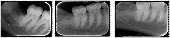
POSTOPERATIVE RADIOGRAPHS AFTER PRP TREATMENT FIGURE 11: 1 MONTH FOLLOW UP, FIGURE 12: 3 MONTHS FOLLOW UP, FIGURE 13: 6 MONTHS FOLLOW UP
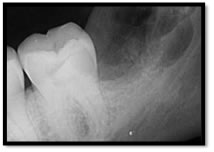
POSTOPERATIVE RADIOGRAPH WITHOUT PRP
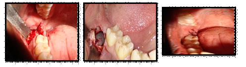
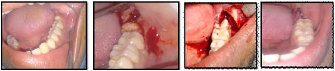
INTRAOPERATIVE PICTURES WITHOUT PRP Fig.14: INTRAORAL PICTURE Fig.15: INCISION and FLAP REFLECTION Fig. 16: SOCKET AFTER REMOVAL Fig.17: SUTURING DONE OF TOOTH RESULTS Total 40 patients were included in the study out of which 20 each were there in both group 1 without PRF includes 10 patients of age ranging in between 20-30 years, and 8 patients from age range 30-40 and 1 each in age range of 40-50 and above 50 respectively. Where as in with PRF group out of 20 patients, 9 patients were belonging to 20-30 years and 7 were there in 30-40 age group,3 in 40-50-year age group and 1 was there in above 50 age group.
TABLE 1: AGE DISTRIBUTION
TABLE 2: SITE LEFT/RIGHT:
Out of 40 patients in without PRF group 10 patient each in which right and left side mandibular mesioangular third molar was removed and in without PRF group 12 in right side and 8 were in left side was affected.
TABLE 3: MEAN DURATION OF SURGERY
*p < 0.05considered statistically significant In above table mean duration of surgery was 36.55 + 8.99 minutes whereas in with PRF group mean duration of surgery was 54.40+ 8.02 minutes. The t- value for mean duration of surgery is 6.626 and p value is 0.000* which is statistically significant. TABLE 4: INTERINCISAL MOUTH OPENING IN MM
*p < 0.05 considered statistically significant Preoperative Mean interincisal mouth opening in without PRF group was 42.45+ 12.16 mm and 47.35+ 5.78 mm in with PRF group. And t value was 1.6276 and p value was 0.119.
TABLE 5: PAIN ON VAS SCALE:
*p < 0.05considered statistically significant In above table mean pain measurement on VAS Scale was 4.60+ 1.67 preoperatively in without PRF group and 4.30+ 1.75, and the t- value was 0.555 and p-value was 0.582.
TABLE 6: RESCUE MEDICATION
*p < 0.05considered statistically significant In this table total no. of rescue medication were analyzed the mean no. of rescue medication was 9.80+ 1.67 in without PRF group and 5.60+ 1.57 in with PRF group and a t value was 8.187 and p value 0.00 was obtained.
TABLE 7: SWELLING FROM LATERAL CANTHUS OF EYE TO ANGLE OF MANDIBLE
*p < 0.05considered statistically significant
TABLE 8: SWELLING FROM CORNER OF MOUTH TO TRAGUS
*p < 0.05considered statistically significant
TABLE 9: SWELLING FROM TRAGUS TO SOFT TISSUE POGONION
*p < 0.05considered statistically significant
TABLE 10: BONE DENSITY
*p < 0.05considered statistically significant In this table mean bone density on post-operative 1 month follow up mean bone density was 85.20 + 13.88 in without PRF group and 98.31 + 13.43 in with PRF group the t value was 3.036 and p value 0.0 04 was obtained.
TABLE 11: POCKET DEPTH
*p < 0.05considered statistically significant In this table mean pocket depth preoperatively was 3.50+ 1.57 in without PRF group and 1.70 + 1.53 and t value was 3.674 and p value was 0.001 and on post op 2nd day mean pocket depth was 7.35+ 1.04 in without PRF group and 3.60 + 1.79 in PRF group and the t value was 8.105 and p value was 0.000 obtained. DISCUSSION Third molar surgery is one of the most common operations in oral and maxillofacial surgery.10 Surgical extraction of third molars is often accompanied by pain, swelling, trismus, and general oral dysfunction during the healing period. Careful surgical technique and scrupulous perioperative care can minimize the frequency of complications and limit their severity. Various pharmacological and/or extraction methods have been used for maintaining patient’s social activities. However, the amount and intensity of edema, pain and trismus occurring after surgical extraction cannot be eliminated completely.11 Platelet rich fibrin is a second generation PRP where autologous leucocytes are present in a complex fibrin matrix to accelerate the healing of soft and hard tissue and are used as a tissue engineering scaffold. Bioactive surgical additives has been used to regulate the inflammation and increase the speed of healing process.12 With this background, the present study was designed to evaluate the healing outcome in mesioangular impacted mandibular third molar using platelet rich fibrin. In the present study, 21 to 30-year age group had the highest prevalence of impaction, followed by patients between 31and 40 years. From this study, it is evident that impacted third molars decrease with corresponding increase in the age of patients. Hashemipou et al also observed that more than half of the patients with impaction were in the third decade of their life.13Similar results were found in study conducted by Sadeta et al.13 It could be because, these patients were symptomatic and were convinced for the treatment. Moreover, it may be because of oral health awareness. The present study revealed that right side was more commonly affected than the left side. In contrast to our study, Ramamurthy et al conducted a study and finds higher prevalence of impaction on the left side than right side.14 There was also no significant difference between the right and left sides in a study conducted by Hashemipou et al.13 The reason for the difference could be the fact that the studies were conducted in different demographic area. A statistically significant difference was observed in the duration of surgery between the case and the control group in the present study. It could attribute to the fact that there was consumption of time in preparation of PRF during the surgical procedure. In the present study, interincisal distance was recorded in all patients preoperatively, on the second postoperative day, seventh postoperative day and at 1, 3 and 6 months postoperatively to assess restriction in mouth opening in the case and control group. A statistically significant difference was observed on the second and seventh postoperative day, and at 1 month follow up. There was no significant difference in interincisal distance at 3 and 6 months follow up. Similar results were found in a study conducted by Kumar et al and Uyaniket al.15,16 This difference in the case and control group indicates that the use of PRF influenced the degree of restriction of mouth opening by increases the efficiency of cell proliferation. PRF releases high quantities of three main growth factors, transforming growth factor β-1 (TGF beta-1), platelet-derived growth factor AB (PDGF-AB), vascular endothelial growth factor (VEGF), and an important coagulation matricellular glycoprotein (thrombospondin-1, TSP-1) during 7 days. Apart from these, PRF also secrete EGF, FGF, and three important pro-inflammatory cytokines- IL-1b, IL-6, and TNF-α which stimulate several biological functions such as chemotaxis, angiogenesis, proliferation, differentiation, modulation leading to regeneration of hard and soft tissues. The present study revealed that PRF significantly recorded less pain for the second and seventh postoperative days, whereas no differences were observed for the first, third and sixth postoperative months when compared with case and control group. Al-Hamed et al conducted a similar study and found reduced pain on the fifth, sixth and seventh postoperative days.17 Similar results were observed in studies conducted by Kumar et al and Uyanik et al.15,16 The reason may be, PRF promotes better and faster healing of the extraction sockets as fibrin present in PRF acts as biological additive that regulates inflammation and increases healing thus reducing pain postoperatively. Thus, the present study revealed that the application of PRF after removal of impacted third molar lessens the severity of immediate postoperative sequelae, decreases preoperative pocket depth, and hastens bone formation.
SUMMARY AND CONCLUSION The finding of present study concluded that PRF application after impacted mandibular third molar surgery is a good biologic material to avoid post-operative sequalae like pain, trismus, swelling. We found definite clinical advantages with respect to the healing of soft tissues. As far as bone was concerned, there was an increase in quantity in the experimental group throughout the study period. And regarding pain and trismus we found less trismus and less pain as compared to control group. Thus Platelet-rich fibrin offers a natural way of augmenting soft and hard tissues. The results of our study add weight to those that advocate the use of platelet- rich fibrin.
REFERENCE
|
||||||||||||||||||||||||||||||||||||||||||||||||||||||||||||||||||||||||||||||||||||||||||||||||||||||||||||||||||||||||||||||||||||||||||||||||||||||||||||||||||||||||||||||||||||||||||||||||||||||||||||||||||||||||||||||||||||||||||||||||||||||||||||||||||||||||||||||||||||||||||||||||||||||||||||||||||||||||||||||||||||||||||||||||||||||||||||||||||||||||||||||||||||||||||||||||||||||||||||||||||||||||||||||||||

 This work is licensed under a Creative Commons Attribution-NonCommercial 4.0 International License.