 Home
Home
Official Journals By StatPerson Publication
|
Table of Content - Volume 12 Issue 3 - December 2019
Retrospective study of the clinical and histopathological parameters of the dental lesions
Shilpi Daveshwar1, Hiral Samir Shah2*, Meena Rajiv Daveshwar3
1PG student, Manubhai Patel Dental College and Dental Hospital, Baroda. 2Tutor, 2Associate Professor, S.S.G. Hospital and Medical College, Baroda. Email: hiralsamir07@yahoo.in
Abstract Background: Oral cavity is the site of myriads of lesions that may be categorized as inflammatory/reactive, cystic and neoplastic. The possibility of identifying the frequency of each lesion is fundamental in order to understand disease patterns within populations and support differential diagnosis. The aim of our study was to investigate the frequency and spectrum of oral lesions biopsied in a hospital population in order to provide insight into the prevalence of them. Methods: A retrospective study of 94 specimens received in Pathology Department from Dental OPD from January 2015 to December 2019 was conducted. The histopathology slides and reports were retrieved from Pathology Department. The histopathological diagnosis was divided into categories considering several variables (type of lesion, gender and age of the affected patient). A statistical analysis was conducted in order to evaluate the prevalence for each cluster of pathology and its relationship with the other identified variables. Results: In group 1, there were 29 samples which showed mainly nonspecific inflammation. Group 2, which consisted of total 23 samples of cystic lesions among which radicular cyst was the most frequent diagnosis. Maximum number of lesions were in group 3 which consisted of 42 neoplastic diseases (44.6%). In third group, malignant tumors represented 25.5% (24/94 cases) of the samples followed by benign tumours and premalignant lesions. Males represented 58.5% of the sample, while females were 41.5%. The overall male to female ratio was1.4:1. The patients’ age range was from 2 to 79 years and mean age was 34.9 year. Conclusions: At present time, the most reliable diagnostic tool is represented by histopathological analysis, which is essential in terms of morphological characterization of the lesion and still remains the gold standard for obtaining a definitive diagnosis. Many of our findings are consistent with those reported in literature, suggesting that the study of demographic characteristics and their association with occurrence of lesions should be considered in performing differential diagnosis. Key Words: Biopsy, Dental Lesions, Histopathological Examination.
INTRODUCTION The word biopsy originates from the Greek terms bios (life) and opsis (vision): vision of life. Biopsy is defined as “The removal of tissue from a living person for microscopic examination to confirm or to establish the diagnosis.” 1The term was coined by Ernst Henry in 1879. The possibility of identifying the frequency of each lesion is fundamental in order to understand disease patterns within populations and support differential diagnosis. According to the American Academy of oral and Maxillofacial Pathology, any abnormal tissue removed from the oral region should be submitted to pathologist for final histopathological diagnosis. The exceptions are in cases such as tori, exostosis, carious teeth lacking attached soft tissue, extirpated dental pulp and clinically normal tissues.2 It is important for the clinician to decide whether a lesion needs to be biopsied or not before treating it. With regard to oral soft tissues, any lesion in question, if persisting for more than 2 weeks even after the removal of the irritating factor (if any), biopsy should be performed. Biopsy is also advisable in bony lesions that cannot be diagnosed radiographically and which are usually accompanied by pain, sensation alterations or other symptoms3. Biopsy has been used since more than 150 years to establish the diagnosis of an unknown medical condition.4 It is widely used in the medical field, but the practice is not quite widespread in dental practice. Biopsy is one of the oldest, gold standard and most reliable currently available method that can establish the definitive diagnosis of clinical abnormalities. The diagnosis of dental lesions is established from the different clinical and radiological features though the final diagnosis is based on histopathological examination of the lesion.5,6 Thus Oral biopsy is considered essential for i) To establish a definitive diagnosis as early as possible so that correct treatment may be initiated without delay, ii) to establish a prognosis in malignant or premalignant lesions iii) To determine whether an abnormality has been completely removed, iv) act as a document with medico legal value. Failure to diagnose oral disease may have profound implications for both the patient and the dentist. Although absolute contraindications are not present, there are some conditions where decision to proceed with biopsy should be done with caution. These are bleeding diathesis secondary to anticoagulation, lesion located near vital structures that could be injured by biopsy and in medical conditions that do not allow for the use of local anesthetics. Biopsy is not advised in the case of multiple neurofibromas due to the risk of neurosarcomatous transformation, or in tumors of the major salivary glands. Such biopsies must be performed by specialized surgeons in order to avoid damaging the nearby anatomical structures and causing the spread of tumor cells, as this would adversely affect the prognosis.3 Aim of present study is to classify and determine the frequency of distribution of various inflammatory, cystic, preneoplastic and neoplastic dental lesions received for histopathological examination from dental department at tertiary care center. Final histopathological diagnosis was given in each case in correlation with the clinical findings.
METHODS This retrospective study was performed at histopathology section of Pathology department, SSG Hospital and Medical College, Baroda. The biopsies of oral lesions were received from dental department of SSG Hospital. The patient records of histopathology sections from January 2015 to December 2019 were reviewed considering gender, age and histopathological diagnosis. Lesions were divided into three major categories based on their histopathologic diagnosis: Group 1: Inflammatory/reactive oral lesions, Group 2: Cystic lesions and Group 3: Benign, premalignant and malignant lesions.7,8The results obtained were tabulated and analyzed. Statistical analysis All results were analyzed using aspects of descriptive statistics. The data was analyzed in different age groups and gender by using frequency, ratio and percentage methods. For evaluation of association between mean age group and number of cases, χ2 test was applied. Significance was considered for values of p<0.05.
RESULTS Total 94 cases were studied and classified in three groups. Distribution of cases among these three groups were analyzed. In Group 1 there were 29cases (30.9%), group 2 had 23cases (24.5%) and group 3 consisted of 42 cases(44.6%). In view of gender distribution, 55cases (61.2%) were males and 39(38.8%) cases were females. The overall male to female ratio was 1.4:1. The patients’ age range was from 2 to 79 years and mean age was 34.9 year (Table 1). Group 1 included inflammatory or reactive conditions, had mean age of 35 year with M:F ratio 1.4:1. Histopathological examination showed 17 cases (58.6%) of chronic inflammatory cell infiltration along with other specific inflammatory lesions like mucormycosis, osteomyelitis, pericoronitis, pyogenic granuloma, inflamed epulis, fibroepithelial polyp and submucosal fibrosis. Group 2 included cystic lesions, with mean age of 24.5 year and M:F ratio was 1.5:1. In this group, the most prevalent was radicular cyst (n=8, 34.7%), followed by nasolabial cyst (n=4, 13.04%). Group 3 consisted of majority cases; including benign, premalignant and malignant lesions. Here mean age was 42.4 year and M:F ratio was 1.3:1. Among benign lesions, Fibroma and its subtypes and ameloblastoma were the most common tumors along with a each case of inflammatory myofibroblatic tumor, adenomatoid odontogenic tumor, lipoma, neurofibroma and psuedoepitheliomatous hyperplasia. Each case of verrucous hyperplasia and carcinoma in situ were included as premalignant conditions. Both were male with mean age of 50 year. Among malignant lesions, squamous cell carcinoma (n=20, 47.6%) was the most common tumor along with two cases of intraosseous mucoepidermoid carcinoma(n=2,4.8%) and each case of plasmacytoma (n=1,2.3%) and oral malignant melanoma(n=1, 2.3%).
Table 1: Shows Frequency of Various Lesions according to Number of Cases, Gender, Mean Age and Histopathological Diagnosis
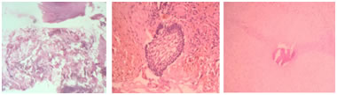
Figure 1 Figure 2 Figure 3 Figure 4 Figure 5 Figure 6 Figure 7 Figure 1: Mucormycosis Showing Large, Nonseptate, Branching Fungal Hyphae. Tissue Necrosis is seen as it is Angioinvasive.(HandE Stain 400X); Figure 2: Amelolastoma Showing Odontogenic Epithelial Islands with Peripheral Palisading of Columnar Cells and Stellate Reticulum like Cells.(HandE Stain 400X); Figure 3: Hypercellular Fibrous Connective Tissue within which Irregularly Shaped Calicifications suggestive of cemento-ossifying fibroma (HandE Stain 100X); Figure 4: Carcinoma in Situ showing Severely Dysplastic Squamous Epithelium with Intact Basement Membrane .(HandE Stain 400X); Figure 5: Plasmacytoma showing Monomorphous Population of Plasma Cells having Eccentric Placed Nucleus and Abundant Cytoplasm. (HandE Stain 400X)(Inset Shows CD 138 Positivity on Immunohistochemistry); Figure 6: Mucoepidermoid Carcinoma showing Cords and Sheets of Mucous Cells, some clear cells and Nests of Squamous Cells. Inset Shows Extracytoplasmic Mucin. (HandE Stain 400X); Figure 7: The cells are highly pleomorphic in nests with dense intra and extra- cytoplasmic melanin pigment. Inset shows positivity for Melan A confirms the diagnosis of malignant melanoma. DISCUSSION It is an accepted fact that microscopic analysis is the gold standard for the diagnosis of most lesions. Present study demonstrate general profile of oral lesions and attempts best to report and classify them. It is based on histopathological examination of oral lesions in correlation with clinical findings to predict significant level of patients’ prognostic outcome. It was difficult to compare the results with other studies as they were performed in a specific group of lesions9,10 and age.11 Teye et al studied 638 jaw swellings and broadly categorized as benign tumors, cysts, infective lesions and malignant neoplasms. M: F ratio was 1.1:1. Mean age was 32.36 ± 17.14 years with a range of 3 to 84 years. Ameloblastoma was the commonest odontogenic tumor followed by fibromyxoma.7 Elif Pecker et al studied jaw lesions for correlation of clinical and histopathological diagnosis. In this study the lesions were divided into three groups that is inflammatory/developmental lesions, cystic lesion and tumor and tumor like lesions. Here distribution of lesions in each group was 26.9%. 53.6% and 19.5% respectively. Mean age was 40 ±1.9 years.8 We followed the same simple and most acceptable way of classification for easy implementation of treatment and prognosis part for clinician. Typically inflammatory swellings are expected to be the type of oral lesions that are caused by mechanical and chemical trauma, radiation injury, infections and immunological mechanisms. In our study they formed 30.9% of all oral lesions. Occurrence of this group of lesions is little more frequently seen in males(1.4:1) as seen in this study and was also supported by Mosby et al.12 Alveolar bone mucormycosis is rare that frequently occurs after tooth extraction in immunocompromised patients and requires aggressive clinical management(Figure 1).13Osteomyelitis of the jaws also occur in immuno-compromised patients both locally and generally following dental treatment. It is a rare condition in jaw characterized by exposed bone in mouth which fails to heal after appropriate intervention.14 The embryonic "resting" epithelium (Also termed cell rests) is usually dormant or, undergoes atrophy but when stimulated may be due to inflammation, may form a cyst.(15) Within group 2 lesions, radicular cysts were the most prevalent cyst(n=8, 34.7 %) followed by nasaolabial cysts(n=4,17.4 %). A greater incidence was in males than in females(1.5:1) which was confirmed by conclusion of other study.(16)Radicular cysts were the most biopsied lesions followed by dentigerous cysts and residual cysts in cystic lesions and these data support the data presented by Nuñez Urrutiaet al.17 The cyst lining Epithelium may be derived in some case from 1) Respiratory epithelium from maxillary sinus when the periapical communicates with the maxillary sinus 2) Oral epithelium from a fistulous tract 3) Oral epithelium proliferating apically from a periodontal pocket. Their incidence is highest in third and fourth decade of lifes with male dominance.18 Third major group includes benign, preneoplastic and malignant lesions(n=42, 44.6%). The most frequent lesion included in benign category was fibroma, a benign neoplasm of fibroblastic origin, is reactive in nature and represents a reactive hyperplasia of fibrous connective tissue in response to local irritationor trauma rather than true neoplasm.19 Ameloblastoma is a benign tumor that is “usually unicentric, nonfunctional, intermittent in growth, anatomically benign and clinically persistent.” About 80% of ameloblastomas occurs in the mandible.(20)The World Health Organization (WHO) (1991) defined ameloblastoma as a benign but locally aggressive tumor with a high tendency to recur, consisting of proliferating odontogenic epithelium lying in a fibrous stroma (Figure 2).21 The main modality of treatment is surgery, with wide resection recommended due to the high recurrence rate. Cementm forming perioodontogenic tumor, cementossifyning fibroma showed histologically hypercellular fibrous connective tissue within which was irregularly shaped calicifications.(Figure 3) The recommended treatment is enucleation of smaller ossifying fibromas, curettage of lesions where no clear radiolucency is present around the lesion and mono-bloc resection with bone reconstruction for larger tumors in close proximity to the inferior border of the mandible.22 Despite the easy access of oral cavity for direct visual examination and advances in treatment for Oral Squamous Cell Carcinoma, the 5-year survival rate remains at <60%.23 Identification of premalignant lesions or early carcinoma with intensified follow up assessments is an ideal strategy to improve survival rates. The premalignant and malignant lesions are closely related lesions which cannot be diagnosed clinically. Therefore, the diagnosis must be established histopathologically. Accurate histopathologic diagnosis depends on an adequate depth of the biopsy specimen and the adjacent normal epithelium.24Present study also includes two rare preneoplastic lesions that are verrucous hyperplasia and carcinoma in situ (Figure 4). Plasmacytoma is a disorder characterized by uncontrolled monoclonal proliferation of plasma cells that causes overproduction of immunoglobulins in blood (Figure 5).25 Plasmacytoma is clinical entity where it is very difficult to confirm the diagnosis without radiological, histopathological, immunohistochemical and other supportive investigative modalities. In our case radiological findings were correlated with histopathological findings and for establishing the final diagnosis confirmed by Immunohistochemistry (IHC) positivity for CD138.Mucoepidermoid carcinomas infrequently arise centrally within the mandible as in our two cases(Figure 6).They usually arise in the salivary glands or sinonasal cavities. On radiograph, mucoepidermoid carcinoma in the jaw typically appear more infiltrative.26 Intraosseous mucoepidermoid carcinoma is more common in middle-aged adults and have a slight female predilection. It is three times more common in the mandible than in the maxilla and are most often found in the area of the molars and mandibular ramus. The most frequent symptom is cortical bulging, although some lesions may be discovered as an accidental finding on radiographs. The main modality of treatment for patients with this neoplasm is radical surgical resection, offering a greater chance of cure than the more conservative procedures, such as enucleation or curettage, that have great possibility of recurrence and tumor metastasis. Oral mucosal malignant melanoma is rare representing about 0.5% of oral malignancies and less than 0.01% of all oral biopsies(Figure 7). It arise in adults with an average age of about 55 years. Though surgical excision is the gold standard treatment, it may be combined with chemotherapy, radiotherapy and immunotherapy27.The malignant lesions are The global burden because of the aging and growth of the world population alongside an increasing adoption of cancer-causing behaviors like smoking, betel quid chewing and alcohol consumption.28Oral Cancer is the 6th most frequent malignant tumour.29There are nearly 300 Dental Collegesin all over the India. Despite number of Dental surgeon passing out from so many Dental institutions, Still India stands first among high incidence of oral cancer cases. In support of present study, Monika et al also proves the maximum number of cases of malignant lesions. 16
CONCLUSIONS The diagnosis of dental lesions should be based on clinical, radiographic and histopathologic features. Oral and Maxillofacial Surgeons must establish the histological diagnosis of their cases by routine biopsy and provide an adequate treatment, which might involve further procedures. This will prevent unnecessary treatments, delayed surgical operations and disease related mortality.
REFERENCES
|
||||||||||||||||||||||||||||||||||||||||||||||||||||||||||||||||||||||||||||||||||||||||||||||||||

 This work is licensed under a Creative Commons Attribution-NonCommercial 4.0 International License.