 Home
HomeOfficial Journals By StatPerson Publication
|
Table of Content-Volume 5 Issue 1 January 2018
Aesthetic replacement of anterior teeth using ovate pontic – A Case Report
Sakshi Anand1*, P Laxman2
1Senior Lecturer, 2Professor and HOD, Department of Prosthodontics, Army College of Dental Sciences, Secunderabad, INDIA. Email: sakshianand26@yahoo.co.in
Abstract Replacing lost teeth aesthetically has always been a challenge for a Prosthodontist. It is very important to preserve the interproximal tissues after the extraction of tooth/teeth for a good aesthetic outcome. After extraction of the tooth, there is recession of the interproximal papilla and collapse of the buccal plate. This makes it difficult to restore the aesthetics to full satisfaction. Hence it is important to preserve the socket size, shape and the space of the gingival tissue in order to preserve the tissue height. This article discusses the successful creation of natural gingival profiles of missing anterior teeth by using ovate pontic. Key Word: Aesthetics; interproximal papilla; ovate pontic.
INTRODUCTION The ovate pontic design is commonly used to maintain or enhance the soft tissue contours of fixed partial dentures.1 Its convex design has been recommended to fulfil the esthetic, functional and hygienic requirement.1,2 Although ovate pontics have been placed in posterior or anterior quadrants with equal success, it is essential to establish exact dimensions, and to condition the gingival tissue underlying ovate pontics early by using a provisional restoration with a convex ovate shape, which acts as a template.1,2 Moreover, since surface roughness results in a simultaneous increase in plaque accumulation, thereby heightening the risk of periodontal inflammation, smoothness of the pontic materials is advised to maintain healthy tissues.3
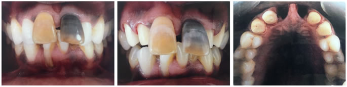
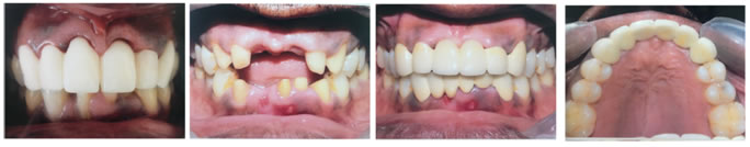
CASE REPORT A 42-year-old healthy male patient reported to the Department of Prosthodontics, Army College of Dental Sciences, Secunderabad, dissatisfied with the appearance of his upper front teeth. Initial evaluation of patient included a detailed social, dental and medical history. On intraoral examination, 11 and 21 were found to be discoloured and proclined. Severe crowding was observed in lower anteriors. (Figure 1) The patient was concerned about his appearance and smile. Patient had good oral hygiene and was caries-free. 41 and 42 were periodontically compromised. He also had a high lip line. Therefore it was decided to extract the upper central incisors and replace them with ovate pontics which exactly simulates the lost teeth in form, function and aesthetics. The patient was informed about the procedure, its cost, the expected clinical longevity, the time period necessary to conclude the treatment, and the possible aesthetic results. Informed consent was obtained from the patient and treatment was initiated. LEGENDS Figure 1: Pre-operative intra oral view of the patient with teeth in occlusion; Figure 2: Biomechanical tooth preparation of teeth adjacent to the teeth to be extracted; Figure 3: Intra oral view after the extraction of central incisors; Figure 4: Provisional fixed partial denture with tissue surface of the ovate pontic 2-3 mm inside the socket; Figure 5: Intra oral view of the extraction socket after 3 months of healing period; Figure 6: Postoperative view of restored maxillary and mandibular teeth; Figure 7: Postoperative maxillary occlusal view of the restoration PROCEDURE
DISCUSSION Ever increasing demands of patients for natural appearing artificial teeth has led to the development of various treatment modalities to achieve natural appearance of soft tissues around the replaced teeth.5 Among these, use of ovate pontic is one of the most versatile and effective means for obtaining desired aesthetics.6 It was first developed by Abrams in 1980.7 The concept has been successfully used in conjunction with fixed and removable partial dentures and also with implant prosthesis. The tissue surface of the ovate pontic should be kept clean using super floss to prevent possible tissue inflammation. The histological study of the tissue surface of the pontic has not revealed any clinically significant problem with this design when appropriate oral hygiene is maintained.8 Hence proper oral hygiene regimen should be followed with dental floss to provide continuous moderate pressure against the apex of the pontic. This procedure has some disadvantages as well. Tissue healing under provisional restorations with ovate pontics is time-consuming. Detailed evaluation of the existing provisional restoration and also the tissue healing is important for an acceptable marginal fit and good aesthetic outcome. Final elastomeric impressions for the FPD should be made immediately after removal of the provisional restoration to prevent tissue rebound.
CONCLUSION Considering the limitations of the observational period, it was observed that an ovate pontic design can achieve aesthetic, functional and hygienic requirement of an artificial tooth. It is recommended for replacing missing teeth particularly in aesthetic zone and in patients with high smile line. The ovate pontic creates an illusion of free gingival margin and interdentalpapilla minimizing the black triangles. Also, maintenance of good oral hygiene by the patient is critical for a successful outcome. Having an excellent working relationship with a dental laboratory is highly recommended.
REFERENCES
|
 This work is licensed under a Creative Commons Attribution-NonCommercial 4.0 International License.