 Home
HomeOfficial Journals By StatPerson Publication
|
Table of Content-Volume 5 Issue 2 February 2018
Zirconia abutment for single-tooth implant – A Case Report
Sakshi Anand1*, P Laxman2
1Senior Lecturer, 2Professor and HOD, Department of Prosthodontics, Army College of Dental Sciences, Secunderabad, INDIA. Email: sakshianand26@yahoo.co.in
Abstract Titanium implant abutments have been the most widely used material for many years as they are biocompatible and provide excellent mechanical retention. However, in clinical situations wherein there is thin periodontal biotype or an unexpected soft tissue recession around the implant, the metallic grey colour becomes evident, becoming an aesthetic failure. Hence, there is a shift from titanium abutment material to zirconium abutment material which has high translucency and mechanical strength. This article discusses the aesthetic replacement of anterior tooth of a young girl using zirconium implant abutment. Key Words: Titanium implant abutment; periodontal biotype; aesthetic; Zirconium Implant Abutment.
INTRODUCTION Over the years, successful osteointegration and function have been the main goals of implant dentistry.1, 2 Implant survival rates have reached extremely high number, however, success of single-tooth implants is no longer defined solely by the survival of the implant. Because of the ever increasing demands of the patient, the focus is now shifting towards aesthetic parameters also.3 Exact replication of the natural dentition, maintenance of a harmonious soft- and hard-tissue architecture,4,5 and invisible integration of the final implant prosthesis are among the challenges of modern implant dentistry.6 Titanium and other metal alloy implant abutments are biocompatible and have provided reliable substructures for implant/abutment-supported crowns. But often their grey metallic colour leads to grey or blue discolorations of soft tissues around the implant.7 Moreover, recession of peri-implant soft tissue may lead to exposure of the metal abutment, compromising the appearance of an otherwise aesthetically pleasing restoration. Zirconium implant abutments were introduced in implantology in an effort to overcome this disadvantage. When compared to titanium abutments, these abutments possess excellent biological advantages also such as low bacterial adhesion and biocompatibility with soft tissue.
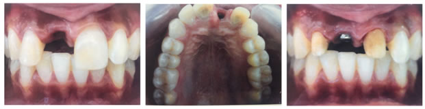
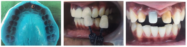
CASE REPORT A 24-year-old female patient with apparent good health reported to the Department of Prosthodontics, Army College of Dental Sciences, Secunderabad, with a missing upper front tooth for the past 6 months. Initial evaluation of patient included a detailed social, dental and medical history. Intraoral and radiological examination revealed endodontically treated maxillary right lateral and left central incisor (Figure 1). The patient was concerned about her appearance and smile. Patient had good oral hygiene and was caries-free. After discussing different treatment options with the patient, cost, time period required to conclude the treatment and the possible aesthetic result, it was decided to place implant in the area of missing central incisor and zirconia crowns on endodontically treated teeth. Informed consent was obtained from the patient and treatment was initiated.
   x x
Figure 1 Figure 2 Figure 3
Figure 4 Figure 5 Figure 6
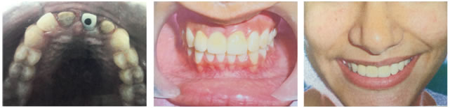
Figure 7 Figure 8 Figure 9 LEGENDS Figure 1: Preoperative intraoral front view showing missing maxillary right central incisor; Figure 2: Intra oral view after placement of implant in maxillary right central incisor; Figure 3: Intraoral front view of the prepared natural teeth and the Healing abutment on Implant; Figure 4: Shade matching for definitive restorations; Figure 5: Elastomeric impression of the maxillary arch; Figure 6: Intraoral front view of the prepared natural teeth and the zirconium abutment before final cementation; Figure 7: Intraoral occlusal view of the prepared natural teeth and the zirconium abutment before final cementation; Figure 8: Postoperative intraoral front view of the zirconium crowns on Maxillary left central and maxillary right central and lateral incisor; Figure 9: Postoperative smile
PROCEDURE
DISCUSSION There is increased worldwide acceptance of zirconia implant abutments mainly due to its aesthetic implications. They elicit an excellent chromatic response when translucent, metal free restorations are luted onto them and also titanium is not exposed when there is soft tissue recession. Also, greyish appearance exhibited by titanium abutment through soft tissue is not seen in zirconium abutment. However, it is observed that when the thickness of mucosa covering zirconium or titanium abutments exceeds 2mm, the difference in colour and light reflection is not noticeable to the human eye.7 Both zirconium implant abutment and titanium implant abutment provide good biological responses.8, 9 From in vitro tests, the main significant data show that the type of connection influences the mechanical strength of zirconia abutments, in that a superior structural resistance can be achieved by means of internal connection via a secondary metallic component; moreover, they show that the use of a secondary metallic component might have a beneficial influence on the stability of zirconia abutments, and that the use of implant-prosthetic zirconia abutments in the molar area is not recommended.10,11,12 Although the strength of both tested systems seems to be adequate to resist physiologic chewing forces in the premolar area 13, zirconia abutments show significantly lower fracture strength than titanium abutments.14 Based on these results, Foong et al.15 said that that one-piece zirconia abutments exhibited a significantly lower fracture resistance than titanium abutment. The mode of failure is specific to the abutment material and design, with the zirconia abutment fracturing before the retentive abutment screw.
CONCLUSION Considering the limitations of the observational period, it was observed that the type of material used for abutment and mucosa thickness bear significant influence on the soft tissue surrounding the implant and subsequent aesthetics. Zirconia abutments will become more popular in implant dentistry for its white colour, differently from metal abutments, for its high level of biocompatibility and for its superior mechanical properties. Moreover, having good working relationship with dental technician and educating the patient about proper oral hygiene maintenance plays a major role in the success of the treatment.
REFERENCES
|
 This work is licensed under a Creative Commons Attribution-NonCommercial 4.0 International License.