 Home
HomeOfficial Journals By StatPerson Publication
|
Table of Content-Volume 7 Issue 1 - July 2018
Management of oral submucous fibrosis by combined oral medication and intralesional injection: A treatment strategy
Shrinivas1, Shushma G2*, Ramalingeshwara Kantly3
{1Assistant Professor, 2Resident, Department of Dentistry} {3Assistant Professor, Department of General Surgery} KIMS, Koppal, INDIA. Email: drshushmamattad@gmail.com
Abstract Background: Oral submucous fibrosis (OSMF) is often described as a condition which is easy to diagnose, but difficult to manage. It is a chronic, debilitating disease of the oral cavity. Though it has a global distribution, it is predominantly a disease of the Indian subcontinent and South East Asia. A standard treatment to manage this condition is not yet available. This article aims at enumerating one of the various treatment modalities of OSMF. Materials and Methods: A total of 30 patients diagnosed with OSMF were treated in Koppal Institute of Medical Sciences for a time period of 9 months, by obtaining the patient’s consent and with the approval of the institution’s research ethical committee. They were treated by administering an intralesional injection of placentrex 1.5ml, dexamethasone1.5 ml, hyaluronidase 1500 IU with 0.5 ml lignocaine HCL injected intralesionally biweekly for 4 weeks along with tablet composition ‘Lycopene, Betacarotene, Selenium, Zinc Sulphate, Copper, Alpha Lipoic Acid’ twice a day followed by mouth opening exercises regularly. Results: Patient’s mouth opening improved for about 7 ± 2 mm (93%), the range being 5-9 mm. Patients had reduced symptoms such as burning sensation, painful ulceration and blanching of oral mucosa. Conclusion: Injection of placentrex, hyaluronidase with dexamethasone combined with oral tablet composition ‘Lycopene, Betacarotene, Selenium, ZincSulphate, Copper, Alpha Lipoic Acid’ is an effective method of managing early stage of OSMF. Key Words:Oral submucous fibrosis, multivitamin, intra-lesional injection.
Oral submucousfibrosis remains enigmatic and characterization of its pathogenesis is still met with difficulties due to the inability to prove a cause-effect relationship in ascribing any known agent in its causation. The fibrogenic potential of areca- nut alkaloids and tanning had been studied extensively and was proved to have an effect in its causation, backed with epidemiological and experimental evidences. A variety of aetiological factors including capsaicin, betal nut alkaloids, hypersensitivity, autoimmunity, genetic predisposition (HLA-A 10, DR3, DR7 and halotypes A10 / DR3, B3 / DR3 and A10 / B8) and malnutrition have been suggested by various authors [1]. It is potentially malignant disorder and crippling condition of oral mucosa. It manifests as blanching and stiffness of the oral mucosa, trismus, burning sensation in the mouth, reduced mobility of the soft palate and tongue, loss of gustatory sensation, intolerance to eating hot and spicy foods and occasionally, mild hearing loss due to blockage of Eustachian tube2. The widely accepted definition of OSMF was proposed by Pindborg and Sirsat in 1966. Oral Submucous Fibrosis is defined as “An insidious chronic disease affecting any part of the oral cavity and sometimes the pharynx. It is always associated with juxta-epithelial inflammatory reaction followed by fibroelastic changes of the lamina propria with epithelial atrophy leading to stiffness of the oral mucosa and causing trismus and inability to speak”3.Worldwide, approximately 2.5 million people are affected, with most cases concentrated on the Indian subcontinent, especially southern India4. Male population affected more than female with ratio of 9.9:15.
MATERIALS AND METHODS This study was conducted on 30 patients with early stages [Grade 1 to 3] OSMF who attended as outpatients in the Department of Dentistry, Koppal Institute of Medical Sciences, Koppal for a span of 9 months. This study was carried out after obtaining the patient’s consent and with the approval of the Institution’s Research Ethical Committee. Clinical diagnosis of OSMF was based on symptoms of burning sensation in the mouth on consumption of spicy or hot food, dryness of mouth, presence of vesicles oral ulcers in the mouth, and restriction of mouth opening were observed [Image 1]. Patients who were medically compromised and those who received previous treatments were not included in the study. Patients presenting in later stages of OSMF [Grade 4, 5 and 6] are not included in our study. The patients were informed about the condition and its precancerous potential and were instructed to quit the use of are canut with tobacco. Their history of personal habits with regard to frequency of chews, duration of use and symptoms like burning sensation and mouth opening were recorded. Out of 30 patients [Grade 3 of OSMF], the number of male subjects was 18 and female subjects were 12. The burning sensation was assessed using visual analogue scale marked from 0 to 10 where 0 indicates no burning sensation and 10 indicates maximum burning sensation. Extraorally, the patient’s mouth opening was measured with reference to interincisal points between upper and lower incisor teeth, the maximum mouth opening was assessed with geometric divider and metallic scale. Intraorally, the findings like blanching of oral mucosa, presence of vesicles and ulcers, palpable bands, limitation of tongue movement were observed. The patients were grouped based on their age: 25-35 year (Group I), 36-45 year (Group II), 46-55 year (Group III) and 56-65 year (Group IV). They were treated by administering an intralesional injection of mixture of placentrex 1.5ml, dexamethasone1.5 ml, hyaluronidase 1500 IU with 0.5 ml lignocaine HCL injected intra-lesionally twice a week for 4 weeks along with tablet composition ‘Lycopene, Betacarotene, Selenium, ZincSulphate, Copper, Alpha Lipoic Acid’ twice a day. Patients were advised for mouth opening exercises regularly. Outcome of the treatment was assessed by measuring postoperative improvement in mouth openingeach week and reduction in symptom of burning sensation using a visual analogue scale. Patients were followed up and observed for period of 9 months and data are tabulated. 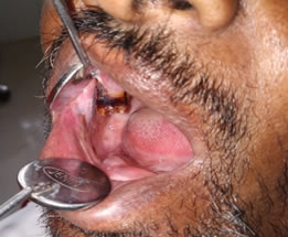 Figure 1: Clinical features of OSMF
RESULTS Improvement in the patient’s mouth opening with a net gain of 7 ± 2 mm (93%), the range being 5-9 mm. Definite reduction in burning sensation, painful ulceration and blanching of oral mucosa was seen in study subjects [Table 1]. It was observed that in Group I prior to treatment, the mouth opening was limited to18%, following the treatment the mouth opening was 31%; the improvement observed was by 13%. In Group II, prior to treatment, the mouth opening was limited to16%, following the treatment the mouth opening was 29%, the improvement observed was by 13%. In Group III, prior to treatment, the mouth opening was 17%, following the treatment, the mouth opening was 30%, and improvement was by 13%. In Group IV, prior to treatment the mouth opening was 19% following the treatment the mouth opening was 28%, the improvement observed was by 9% [Table 2 and Image 2].
Table 1: Relief of symptoms after treatment
Table 2: Average mouth opening before and after treatment.
Figure 2: Mouth opening before and after treatment
DISCUSSION OSMF is one of the precancerous conditions of the oral cavity most commonly seen in Indian subcontinent. This clinical description had been reported since the time of Sushruta6. Pathologically OSMF is considered as metabolic disorder of oral cavity affecting collagen metabolism. Even though OSMF has a multiple factor for its pathogenesis, areca nut chewing habits considered to be main culprit and presence of nutritional deficiency of vitamin B complex and poor oral hygiene will adds up for the prognosis of the disease. Recently the role of microelements like Zinc and Selenium in the integrity of mucosal membrane and collagen metabolism is been in reported. It is believed to involve juxta-epithelial inflammatory response and fibrosis in the lamina propria, possibly due to increased cross-linking of collagen by upregulation of lysyl oxidase activity. This fibrosis, or the upsurge of collagen, results from the property of areca nut, which increases collagen assembly and also decreases collagen deprivation7. In the early stages patients will have burning sensation, blanching oral mucosa, ulceration and xerostomia. Later, the oral mucosa becomes stiff and opaque, by means of fibrous bands on the buccal mucosa, soft palate, lips, and tongue causing limit in mouth opening, intricacy in mastication, speech and swallowing8. Histopathology of the lesions will show epithelial atrophy with loss of rete ridges, epithelial atypia, and pigment incontinence. Lamina propria shows widespread fibrosis of collagen fibres with a chronic inflammatory cell infiltrate. In later stages, subepithelial hyalinization with atrophic changes in minor salivary glands and skeletal muscle can also be seen. Treatment options for OSMF depend on clinical presentation of the lesion and presence or absence of malignant change of the disease. Depending on the clinical features Kakkar and Puri graded OSMF into six grades9. Grade I: Only blanching of oral mucosa without symptoms Grade II: Burning sensation, dryness of mouth, vesicles or ulcer in the mouth without tongue involvement Grade III: In addition of Grade II, restriction of mouth opening Grade IV: In addition to Grade III palpable bands all over the mouth without tongue involvement Grade V: Grade IV and also tongue involvement Grade VI: OSMF along with histopathological proven cancer. Various types of treatment modalities [Table 3] are available for OSMF depending on clinical presentations: conservative methods, medical management, and invasive methods like surgical elimination of the fibrotic bands and combined therapy [10]. Whatever the stage of the disease all the patients require: complete cessation from areca nut chewing, oral physiotherapy and antioxidant supplements. One of the major difficulty in treatment of OSMF is, if, once the disease is evident, it either persists or becomes more severe with contribution of additional areas of the oral mucosa.
Table 3: Different treatment modalities for OSMF:
Quit Areca Chewing Habit and Antioxidant Supplements: Chewing Betel Quid is considered an important etiological factor of OSF because of the excessive reactive oxygen species (ROS) induced by the ingredients of Betel Quid11. Gupta et al in their study found that after 6 weeks of treatment with tablets containing mostly beta-carotene and vitamin E, patients showed an effective increase in mouth opening and tongue protrusion12. Anil and Sharma concluded that either oral zinc alone or in combination with oral vitamin A in grade I and II patients and oral zinc with local cortisone in grade III patients of OSMF may be employed in future to treat OSMF13.Tai et al in their study stated the use of oral administration of milk from cows immunized with human intestinal bacteria which leads to significant improvement of symptoms and signs in OSMF patient14.The various nutritional supplements found to be effective in treating OSMF are: (1) Micronutrients and minerals- Vitamin A, B complex, C, D and E, iron, copper, calcium, zinc, magnesium, selenium and others (2) Milk from immunized cows- 45 g milk powder twice a day for 3 months (3) Lycopene-8 mg twice a day for 2 months. Oral physiotherapy: Stephen and Hans conducted a clinical trial of Physiotherapeutic treatment to improve oral opening in oral submucous fibrosis in the Nepali population and suggested that physiotherapy is effective for increasing the oral opening and can be readily used to improve OSF in communities with otherwise limited health resources15. In our study we used combination therapy including oral supplement with capsule containing Lycopene, Betacarotene, Selenium, ZincSulphate, Copper, Alpha Lipoic Acid and intra –lesional injection of mixture of placentrex 1.5ml, dexamethasone1.5 ml, hyaluronidase 1500 IU with 0.5 ml lignocaine HCL for treating Grade 3 of OSMF. Dexamethasone: They act by their anti-inflammatory activity by inhibiting the generation of inflammatory factors and increasing the apoptosis of inflammatory cells. Thereby partially relieving the patients of their symptoms at an early stage of OSF. Therefore steroids are useful in controlling symptoms, or as an adjunct therapy. Currently, intralesional steroids are the main treatment modality. These are injected into the fibrotic bands biweekly for 6--8 weeks along with mouth-opening exercises8,9. Placentrex: Placentrex is an aqueous extract of human placenta that contains nucleotides, enzymes, vitamins, amino acids, and steroids. It acts by biogenic stimulation and increasing the vascularity of tissues based on the principle of tissue therapy introduced by Filatov in 1933.Katharia reported that placenta extract when administered result in significant improvement in mouth opening, colour of mucosa, burning sensation, and reduction of fibrotic bands [16]. Antifibrotic Intralesional Injections: Hyaluronidase showed a much quicker effect in ameliorating the burning sensation and painful ulceration than did dexamethasone, though the effect was short-term. It acts by depolymerizing hyaluronic acid, which is the ground substance in connective tissue lowering the viscosity of the intercellular cement substance, and decreasing collagen formation [9, 10 and 11].Combination of dexamethasone and hyaluronidase and placenrex gave better long-term results than other regimens with single drug regimen injection therapy17. Study by Borle and Borle postulated that treatment following intralesional injections of various drugs leads to aggravated fibrosis and pronounced trismus6. The resultant worsening of this condition with submucosal injections are attributable to repeated needle stick injuryto the soft tissues at multiple sites, clinical irritation from drugs being injected, and to the progressive nature of the disease18. The same outcome has been observed with some surgical methods employed to treat OSMF. Conservative line of treatment like topical steroids, vitamins, antioxidants, physiotherapy would give expected symptomatic relief of pain and burning sensation19. Kumar et al. study shows that combined therapy employing nutritional and iron supplements with intra-lesional injection therapy using hyaluronidase, dexamethasone and placentrix in addition to local anaesthetic topical gel and topical application of triamcinolone acetonide 0.1% caused a marked improvement in patient’s signs and symptoms. The clinical outcome is evidenced by improvement in colour of the oral mucosa, decrease in blanching and decreased severity of burning sensation, increased mouth opening and tongue protrusion20.
CONCLUSION OSMF is a progressive disease of oral cavity with variety of clinical presentations at different stages of presentation. Combined oral tablets and local injection followed by physiotherapy is safe, cheap and effective in oral submucous fibrosis without any significant side effects and contra indication which gives significant result in early stage [Grade 1 to 3] of OSMF. Quitting of areca nut chewing and oral physiotherapy is must for all patients.
REFERENCES
|
 This work is licensed under a Creative Commons Attribution-NonCommercial 4.0 International License.