 Home
HomeOfficial Journals By StatPerson Publication
|
Table of Content - Volume 11 Issue 2 - August 2019
Endoscopic removal of bullet lodged in left orbit
Shrinivas Chavan1*, Vitthal D Kale2, Vinayak Kurle3, Archana Shyleenderan4
1Professor and HOD, 2Assistant Professor, 3Senior Resident, 4Junior Resident, Department of ENT, Grant Government College, Mumbai, Maharashtra, INDIA. Email: shrinivasc77@gmail.com
Abstract In this era of peace and stringent firearm regulatory norms, the civilian firearm injuries are rarely encountered in the day to day life. These injuries might be resulting from assault, accidental or suicidal attempts. The most common site of lodgement of bullet is maxillary sinus>frontal sinus>ethmoidal sinus> sphenoidal sinus. The lodgement of bullet within the orbit through the forehead narrowly missing the frontal lobe is unheard off. Key Words: Paranasal sinuses, firearm injuries, Endoscopic nasal surgery.
INTRODUCTION The bullet injuries are devastating causing immense physical and psychological trauma to the patient. Bullet injuries to face are often life threatening and demands a meticulous operative management due to the complexity of craniofacial anatomy. Traditionally these injuries were managed by open approach. But with the latest advancements in the endoscopic procedures; endoscopes have been found to be a boon in dealing with these craniofacial skeletal injuries. It not only gives a magnified view of surgical field and but also maintains the cosmesis avoiding a physical and mental scar.
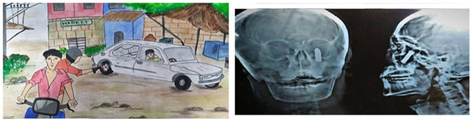
CASE REPORT We are reporting a case of a newly married young man, who sustained a bullet injury, wherein the bullet entering via right side of forehead getting lodged in left orbital apex, miraculously missing the frontal lobe of brain. We hereby detail the methodical approach and endoscopic removal of bullet removal from orbit apex area; without a single scar. Our patient is vegetable vendor; 27 y old male, newlywed, on his way home after completing his daily work. He encountered a typical robbery incident as depicted in the thriller movies. Where two bandits travelling in a two wheeler, parallel to his motor vehicle on the right side, chasing him; one of the bandits in the pillion seat shot our patient, where bullet travelled in a tangential manner hitting the patient’s right side of forehead causing fracture in the frontal part of the cranium narrowly missing the brain and finally lodging in the left orbit. Patient momentarily lost his consciousness and was rushed to the nearby local hospital. He was given preliminary management and other radiological investigations were carried out, which showed the presence of foreign body in left orbital apex. During the course of admission in the local hospital, patient developed slow deterioration of left eye vision, proptosis, chemosis which was progressively worsening every passing day. On the fifth day following the injury the patient came to us for the definitive management. On examination a 1.5 x 0.5 x 0.5 cm wound with scab was noted on the right supra orbital region suggesting the entry point of the bullet. Upon preliminary examination, there was also left eye painful proptosis with chemosis with restricted extra ocular movements. His right eye vision was PL+ and ophthalmology consultation suggested optic neuritis due to long standing exposure to chemicals emitting from the fired bullet. Anterior rhinoscopy showed no specific abnormalities. CT scan showed a cylindrical hyper intense object in the left orbital apex area, fracturing floor of the orbit and the right squamous part of temporal bone without any evidence of cerebral contusion, injury to optic nerve and lamina papyracea. OPERATIVE NOTES Patient was taken under general anaesthesia, and left nostril packed with patty soaked in 4%lignocaine with 1:30,000 adrenaline.30 degree endoscope was introduced into the left nostril, uncinectomy done, left maxillary ostium widened with powered instrument. Blackish colored clots noted in the left maxillary sinus. Blood clots were removed by suction and wash was given into the maxillary sinus with normal saline. Fractured bone chips of orbital floor removed and orbital periosteum was exposed. Orbital periosteum was meticulously dissected out showing presence of golden shining foreign body noted. Soft tissue dissection done creating adequate room for surgery. Firearm bullet was found to be embedded in the orbital tissue, early granulation and fibrous tissue formation. No conventional FESS instruments were used during the procedure. Bullet was initially medialised by hooking with cricoid hook and careful dissection done around the bullet releasing adhesions around it and was finally delivered out via left nostril with the help of Luc’s forceps. Reconstruction of left orbital floor was deferred for second sitting due to the presence of active infection. Packing was done in left nostril with netcell soaked in normal saline after obtaining haemostasis. RESULTS Figure 1: Sketch of the incident(as described by the patient) Figure 2: X-ray skull AP, Lateral view depicting foreign body(bullet) lodged in left orbital apex
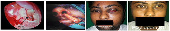
Figure 3 Figure 4 Figure 5 Figure 3: Intra0perative bullet seen embedded in granulation and fibrous tissues; Figure 4: Removal of bullet from left nostril; Figure 5: Pre and post operative images of the patient
DISCUSSION In the past 20 years, endoscopy has emerged as a complement to several standard surgical approaches. Endoscopic techniques which in its initial days were used solely by otorhinolayngologists for functional endoscopic sinus surgery are now widely used for several procedures like CSF leak repair, resection of selected benign and malignant lesions, arriving to extended endoscopic skull base and intracranial procedures.3Foreign bodies in head and neck region mostly involve maxillary sinus of which the most commonly encountered ones are tooth, dental amalgam, gauze piece.4A fired bullet getting lodged in left orbital apex fracturing cranial part of skull narrowly missing the vital cranial structures; managed solely by endoscopic technique, removing the foreign body from orbital apex makes this case truly interesting. Not many cases of foreign body removal in head and neck region are solely managed by endoscopic procedures. Sublabial antrotomy or Caldwell‑Luc approach is the age long procedure used for removal of foreign bodies from maxillary sinuses. This is particularly helpful in case of large or impacted foreign bodies. Bone flap technique may be helpful in case of a large window in Caldwell‑Luc approach. Endoscopic removal1, whenever possible, should be the first choice as it is associated with lesser morbidity and complications and the ease of surgery in well trained hands. Small foreign bodies can easily be removed endoscopically. Endoscopic approach has the advantage of better illumination and visualization. Chances of leftover fragments of foreign bodies are minimum in endoscopic approach. Long standing foreign body can itself lead to complications to vital structures due to the chemicals emitted from them.
CONCLUSION With the advancements in the endoscopic techniques, removal of foreign body lodged in interiors of paranasal sinus, orbital regions can be boldly attempted after having a thorough route plan by imaging procedures like CT PNS. Endoscopic procedures have a better hand over open surgical technique in terms of better illumination, magnification, and cosmetic outcomes. Firearem foreign bodies should be addressed at the earliest; considering adverse effects of long term exposure to chemicals from them like optic neuritis and diminished vision. Fracture of the floor of orbit should be preferably addressed for second stage as it will prevent formation of orbital hematocele.
REFERENCES
|
|
 This work is licensed under a Creative Commons Attribution-NonCommercial 4.0 International License.