 Home
HomeOfficial Journals By StatPerson Publication
|
Table of Content - Volume 12 Issue 2 - November 2019
Chondroid syringoma - Presentation of a case
G M Lucchesi1, G Micali2, F Carfì3, F Asprea4*
1Co- Director of Division of Otolaryngology, 3Senior Consultant, 4HOD, C.O.T Clinic, Via Ducezio, 1, Messina, ITALY. 2Co- Director of Division of Otolaryngology, Head of the Maxillofacial Surgery Unit - C.O.T. Clinic – Via Ducezio, 1, Messina, ITALY. Email: ciccioasprea@gmail.com
Abstract Chondroid syringoma is a mixed tumor of the skin. The histological findings have two origins: epithelial and mesenchymal structures with the tendency to produce chondroid matrix and to differentiate to any form of adnexal structure, mainly sweat glands. It is a solitary, intradermal or subcutaneous tumor, firm and well defined that is located in head and neck. Their behavior is benign and their malignant counterpart is extremely rare. The characteristics of the syringoma chondroid are reviewed and a case is reported. Key Words: chondroid syringoma, mixed tumor of the skin
INTRODUCTION Syringoma chondroid is a rare tumor derived from the major and minor salivary glands. The incidence in primary skin cancer is less than 0.01%. The 80% of chondroid syringomas are observed in old age, commonly in the head and neck area, especially in the gene and nasal region. The authors present a clinical case to discuss the surgical management of these lesions, underlining the importance of including this tumor in the differential diagnosis of head-neck neoformations in the presence of subcutaneous nodules on the face, in middle-aged and male patients. Histopathology. There is no generally accepted classification of the pathological process, however, practical dermatologists to determine the risk level of chondroid siringomy malignancy and timely appointment of adequate therapy using the classification of the disease, aligned with histological features. Based on the presence of flat cells in the chondroid syringoma, V. Lever and G. Schaumburg in 1983 identified two possible variants: 1. The tubular type is characteristic for chondroid siringomy whose elements consist of two layers of epithelial cells - internal plane and prismatic external inclined respect to proliferation and chondroid reproduction matrix, which is capable of regeneration. 2. Cystic type it is characteristic of a single-layer chondroid syringoma in the stroma of which basophils and mucous substances are present, but there are no flat cells, which determine the quality of the tumor. The main elements of the syringoma chondroid are small painless oval pinkish nodules with a shiny surface. Tumor formation is found intradermally or slightly above the surface of the skin. They are similar to pasta to the touch and located where there are sweat glands. Very often the chondroid syringum debuts on the skin of the trunk. The nodules have slow growth, can grow over the years, rarely exceed 5 mm in diameter, sometimes have a translucent membrane, are able to ulcerate. The appearance of primary cells is not accompanied by subjective symptoms or occurrence of expressed aesthetic defect and remain unnoticed for a long time for the patient, which is dangerous in terms of possible syringocarcinoma tumor transformation. Diagnosis. The diagnosis of syringoma chondroid requires mandatory histological confirmation. The presence of epithelial cells and the proliferation of connective tissue with sites of mucinous inclusions and chondroids are detected in the tumor. Apply a bullet or biopsy exam. They use cytology of smear-scarifiers from nodular eruptions (similar to imprints-prints with bladder dermatosis). Acantholytic cells can be detected in the tumor. Differentiate the chondroid syrondum with a mixed tumor of salivary glands, basal cell, syringocarcinoma.
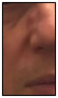
CASE REPORT A married 53-year-old male patient underwent a special consultation at our center due to the presence of a lesion located in the upper part of the nose, right side, characterized from a hemispherical neoformation of about 5 mm of diameter, skin color, smooth surface, firm consistency and mobile on the underlying planes (Figure 1). The evolution is chronic and asymptomatic. The patient also reported that this neoformation had appeared a few years earlier and that over time it had increased in volume, until reaching the actual dimensions.
Figure 1: Clinical appearance of the patient RESULTS After a careful specialist evaluation, the patient was candied for an exeresis of the neoformation. The lesion was removed in its entirety and the surgical region was sutured with aesthetic suture. Histological examination gave a diagnosis of “nodule of adenoid cystic carcinoma”. Subsequently the neoformation was examined by two other centers of pathological anatomy with the final diagnosis of "nodule with net margins and stroma mixo chondroid, mixed tumors of the skin: syringoma chondroid". The patient is asymptomatic and without recurrence.
CONCLUSION We presented a clinical case with exophytic neoformation in the nose with chronic and asymptomatic evolution and whose preoperative diagnoses are part of the differential diagnosis of the syringoma chondroid. Syringoma chondroid is a rare and benign tumor that is difficult to recognize; the diagnosis is always made from histological study and treatment is exeresis. Complete surgical procedure to prevent recurrences.
REFERENCES
|
|
 This work is licensed under a Creative Commons Attribution-NonCommercial 4.0 International License.