 Home
HomeOfficial Journals By StatPerson Publication
|
Table of Content - Volume 12 Issue 3 - December 2019
A study on efficacy of autologous platelet rich plasma usage in myringoplasty
P K Purushothaman1, Preethy Josephine Kennedy2, C R K Balaji3*
1 Professor & HOD, 2PG, 3Associate Professor, Department of Otorhinolaryngology SRM medical college hospital and research centre, SRM Nagar, Kattankulathur, Tamil Nadu-603203, INDIA. Email: preethyjkennedy@gmail.com
Abstract Aim: To compare the effect of autologous platelet rich plasma usage in graft uptake and hearing improvement among patients undergoing Myringoplasty Materials and Method: A prospective study involving 80 patients classified into 40 patient who underwent myringoplasty with autologous PRP usage(Group A) and 40 patient who underwent myringoplasty alone(Group B) was done to assess the graft uptake and hearing improvement postoperatively in the Department. of Otorhinolaryngology, SRM Medical College Hospital and Research from 2018 to 2019.Result: (1) Among the 40 patients who underwent Myringoplasty with Autlogous platelet rich plasma usage , 92.5 % showed graft uptake on post operative day 21 while in Group B only 25 % showed graft uptake. This data was statistically significant( p value of 0.0001). (2) Majority of Group A (95%) did not have any residual or reperforation while group B showed slightly more number of reperforation.(3) Group A showed mean A-B gap improvement of 17.37 (SEM of 0.62) while group B showed mean improvement of 16.75,which was statistically not significant.(4) Infection rate in our study was 2.5 % among group A while 7.5% among group B which was statistically not significant. Key Words: PRP –Platelet rich plasma
INTRODUCTION Autologous platelet rich plasma is gaining popularity world wide and is now being used in majority of the specialities like Oral and maxillofacial Surgery, Dermatology, Orthopaedics, Gynaecology, Plastic surgery, Dentistry, Sports Medicine, Cardiothoracic Surgery since it enhances wound healing due to the growth factors present in it.1 It has shown to influence Human preadipoctyes and tympanic keratinocytes proliferation ,thus playing a main role aiding facial lipostructure sugeries.2 In Face lifting surgeries it was found to improve the edema and ecchymoses post operatively.3 In Musculoskeletal medicine, it has shown to fasten healing process of injuries to tendon, ligament, muscle, bone, nerve and joint/cartilage with good outcomes in pain reduction and restoring mobility.4 In Oral and maxillofacial surgeries such as bone-added osteotome sinus floor elevation with use of platelet rich fibrin as graft material proved to increase the bone formation rate radiologically. In our field of Otorhinolaryngology, platelet rich plasma is being used widely. Most of the intranasal surgeries such as Septoplasty, Endoscopic sinus surgery causes postoperative crusting, bleeding and dryness of nasal mucosa due to disturbance in nasal mucociliary cleacrance.5 The use of Platelet Rich Plasma has shown to increase nasal mucociliary clearance and thus reducing bleeding and crusting postoperatively.5 During Skull base surgeries such as CSF leak repair via endoscopic transsphenoidal approach, leukocyte enriched platelet rich fibrin was launched as a new graft material in endoscopic spontaneous CSF leak repair.6 Otological uses of platelet rich plasma are mainly focused on improving graft uptake during procedures such as Myringoplasty, Tympanoplasty with or without Cortical Mastoidectomy.7It is quoted that inorder to get an intact Tympanic membrane, the success rate is 95 percent .8Many studies have been done internationally which show that Autologous platelet rich plasma increase graft uptake, reduces the healing time, improves hearing, decreases infection rate and improves the overall success rate of getting an intact tympanic membrane postoperatively.7,9,10,11,12,13,14,15Being rich in various growth factors and platelets, it has not only shown to increase the rate of epithelialisation over the graft but also shown to reduce graft displacement significantly due to its adhesive nature.7,9 Some researches also show that it increases the immunity due to which post operative infections are less.7 The purpose of this study was to assess the effect of autologous platelet rich plasma usage on graft uptake and hearing improvement in Myringoplasty .
MATERIALS AND METHODS A randomised controlled study was done in the Dept. of Otorhinolaryngology, SRM Medical College Hospital and Research Centre, on effectiveness of topical autologous Platelet rich plasma usage in Myringoplasty from 2018 to 2019. A total of eighty (80) patients were included in our study . Patients who had Chronic Otitis Media, Tubotympanic or Mucosal type with dry ear for 6 to 8 week after conservative management with oral antibiotics and other supportive measures were included in our study.Figure showing perforation of tympanic membrane pre operatively Patients who were not willing for Myringoplasty were excluded from the study. . They were allotted into two groups: case group included 40 patients who would undergo Myringoplasty with use of autologous PRP and control group included 40 patients who would undergo Myringoplasty without use of autologous PRP. 5-10 ml of blood was collected from patient who were in group A and centrifuged as soft spin and then the sample containing 3 separate layers was taken and top layer consisting of platelet poor plasma was discarded.[16] Buffy coat forming the middle layer was separated alone from the bottom layer of RBC's and transferred into a fresh container and centrifuged as hard spin.Later this sample was taken and the platelet rich plasma was used during myringoplasty to put on the edges of the graft.16
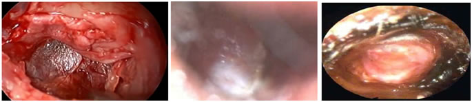
Figure 1 Figure 2 Figure 3 Figure 2: showing graft uptake on post operative day 7 of myringoplasty with autologous platelet rich plasma usage; Figure 3: showing neotympanum on post operative day 21 of Myringoplasty with autologous platelet rich plasma usage Figure showing Intraoperative picture with autologous platelet rich plasma being put at the inferior edge of graft After obtaining informed consent patients underwent Myringoplasty. The results of Otoendoscopic and Microscopic examination were compared between case and control group. Patients were followed every 7th day postoperatively for a duration of 3 months. All statistical analysis were performed using Statistical Package for Social Science (SPSS, version 17) for Microsoft windows. Descriptive statistics were presented as numbers and percentages. The data were expressed as Mean and SD. Chi – square test ,Fischer exact probability test were used for qualitative data. A two sided p value < 0.05 was considered statistically significant. Figure showing Intraoperative picture with autologous platelet rich plasma being put at the inferior edge of graft After obtaining informed consent patients underwent Myringoplasty. The results of Otoendoscopic and Microscopic examination were compared between case and control group. Patients were followed every 7th day postoperatively for a duration of 3 months. All statistical analysis were performed using Statistical Package for Social Science (SPSS, version 17) for Microsoft windows. Descriptive statistics were presented as numbers and percentages. The data were expressed as Mean and SD. Chi – square test ,Fischer exact probability test were used for qualitative data. A two sided p value < 0.05 was considered statistically significant.
OBSERVATION AND RESULTS Out of 80 patients , majority(32.5%) of the cases in group A belong to 31-40 yr age interval while in Group B majority of patients belong to 41-50 yrs age interval. Out of the 40 cases in group A 55% were female while in Group B 52.5 % were males.
Table 1: comparing graft uptake on POD 21
Among Group A patients 92.5 % showed graft uptake on post operative day 21 while in Group B only 25 % showed graft uptake. Chi square test analysis of 37.6 and p value of 0.0001 showed that the data was statistically significant in Group A compared to group B. Table 2: comparing graft uptake on POD 28
By Post op day 28 , Group A showed 95% uptake while only 92.5 % uptake in group B showed graft uptake.On comparing the number of patient with ear discharge among both groups post operatively it was found that in Group A 95 % patients had dry ears while in Group B only 92.5 % showed dry ear. This was statistically insignificant (p>0.05%).
Table 3: showing ear discharge on post operative day 28
Similarly on post operative day 28,Group A showed a slight increase in the number of dry ear patients((97.5%) when compared with Group B which showed (92.5%) but this data was statistically insignificant since p value was 0.3079 (p>0.05).
Table 4: showing reperforation on post operative day 21
With respect to reperforation on 21st and 28th post op day ,Majority of Group A (95%) did not have any residual or reperforation while group B showed slighty more number of reperforation.Chi square anlysis of this data( value of 0.21) revealed no statistical significance. Table 5: showing A-B gap reduction
With respect to A-B gap improvement ,Group A showed mean improvement of 17.37 (SEM of 0.62) while group B showed mean improvement of 16.75. When statistically analysed using chi square test, p value was 0.474 showed no statistical significance.
DISCUSSION El Anwar, 7 in 2015 conducted a randomized controlled trial among 64 patients. They were randomized into case group of 32 subjects for autologous PRP usage in Myringoplasty and control group of 32 subjects for only myringoplasty.7 In this study, age and sex of both groups were statistically matched and the graft uptake in cases (100%) were significantly more than in control group (81.25%) which was statistically significant.7 (P=0.02). A gain in hearing of greater than or equal to ten decibels was noticed among 21 subjects (65.6%) in case group and 11 patients (34.4%) in control group which was statistically not-significant, difference (P=0.079) and rate of Infection in control group (12.5%) was found to be significantly more than in case group (P<0.0001) when analysed statistically.7 When compared with our study which inluded 80 patient, two groups had 40 patients each and 41 of them were females while 39 were males. Graft uptake in cases was seen to be initially 92.5% in case and 25% in control in third week post operatively which improved to 95 % in cases while 92.5% in control study on the fourth week post operatively where as improvement in hearing as well as infection rate between the two groups were statistically not significant. Thus our study was in accordance with their study and concluded that using PRP during Myringoplasty increases closure of perforations in Tympanic membrane , prevents infection and has no side effect .Nithin Prakasan Nair, 9 in 2019 conducted a randomized controlled trial among Eighty-six patients for a duration of two years 43 cases underwent Myringoplasty with usage of autologous platelet rich fibrin while 43 controls underwent Myringoplasty without platelet rich fibrin usage.9 For a period of three months post operatively, patients were followed up and Graft take up rate was around 97.7% in the study group when compared with the control group who showed 81% graft uptake rate which was statistically significant(p=0.012) and 4.7% of the patients in the study group were found to have postoperative infection while in control group only 19% patients had infection after surgery which was statistically significant( p = 0.039).9 The study was similar to our study which also showed 95% graft uptake among cases while 92.5% graft uptake among control study but infection rate in our study was 2.5 % among cases while 7.5% among controls which was statistically not significant and hence not in accordance with their study. Hence comparing both these studies we were able to conclude that platelet rich plasma does have a role in increasing the healing of perforation but whether it plays a role in decreasing infection rate could not be commented. Mehmet Habesolgu , 12 in 2014 did a study on 32 patients with acute Tympanic perforation which showed an increase in graft uptake when PRP was used.12 At the end of the first month after surgery 64.3% closure rate of perforation among cases and 22.2% was the closure rate among controls while towards the last of second month only 1 patient among study group and 4 patient among control group showed failure.12 In our study, out of 40 cases,38 showed complete closure with 2 failed closure. In control study, there were three failures among 40 cases. At follow up after 3 weeks, graft uptake was seen in 92.5 % cases while in the control study25 % only had graft uptake. But after 4 weeks ,95% of our cases had healed tympanic membrane while in the control group 92.5 % had healed perforation. Our study therefore reveals that use of autologous platelet concentrate accelerates tympanic membrane closure. The 2 failure among cases was found to be due to infection. In the control study there were 3 patients whose perforation failed to close. Lyngdoh N C,14 in 2019 conducted an interventional study among 50 patients in the age group of 15-45 years. The success rate in the study was 87.5% and failure rate was 12.5% and that adding PRP to fat myringoplasty had 100% closure rate in small perforation of tympanic membrane and 79.3% closure among medium sized perforation of tympanic membrane whereas the thresholds of air conduction decreased by 9.375 dB post-operatively.14 Our study among 80 patients in age group of 18 -.70 years revealed 95% successful uptake of graft among cases with 5% failure but fat was not used along with platelet rich plasma and air -bone conduction gap reduction was 16.75 after surgery among the cases. When the hearing improvement among cases (17.37) were compared to the control study(16.75) there was no big difference and statistically insignificant (p=0..474). These results were in accordance with their study hence we were able to come to the same conclusion that platelet rich plasma increases the uptake of the graft but whether hearing improves with usage of platelet rich plasma is controversial. Sharma D, 10 in 2018 published a prospective study among 50 patients which were divided into 25 cases who underwent Myringoplaty with autologous platlet rich plasma and 25 controls who underwent Myringoplasty alone.10 Follow up done at the following time intervals revealed that during the first month end 72% of the cases and 40% of the controls showed closure of perforation in Tympanic membrane whereas second month end showed 92% of cases and 70% of controls showed closure of perforation in Tympanic membrane while at the third month end 96% of the cases and 80% of controls showed closure of perforation in Tympanic membrane.10 In our study of 80 patients which were divided into 40 in each group we were able to appreciate graft uptake of 92.5% and 25% in control in third week post operatively which increased to 95 % in cases while 92.5% in control at the fourth week post operatively. Hence both the studies concluded that with platelet rich plasma usage in Myringoplasty, there is an accelerated closure of perforation. Maria Luisa Navarett Alvaro, 15 in 2011 did a pilot study on 3 cases which showed 100% closure of perforation in Tympanic membrane when platelet rich plasma was used in type 1 Tympanoplasty. Even our study majority of the cases (95%) showed graft uptake and complete closure of perforation compared to the control group(92.5%) by the end of fourth week post operatively Hence both studies had similar results showing better success rate and overall outcome after platelet rich plasma was used during Myringoplasty. Ruta Shanmugam, 11 in 2016 conducted a prospective study in which twenty patients underwent Myringoplasty with usage of autologous platelet rich plasma.11 Post operatively recorded all patients showed graft uptake following Myringoplasty with usage of platelet rich plasma and 85 % of the cases showed improvement in hearing of 10 dB.11 This was similar to our study which showed 95% graft uptake among those who underwent Myringoplasty with the usage of autologous platelet rich plasma while 92.5 % graft uptake was appreciated in those who underwent Myringoplasty alone and all the cases showed improvement in hearing of 10dB.11 Thus both studies concluded that platelet rich plasma enhances post operative graft uptake due to presence of growth factors which help in biostimulation.11
CONCLUSION In Myringoplasty when autologous platelet rich plasma is used, it hastens the epithelialisation over the graft leading to faster graft uptake. Since it has a rich platelet concentrate it decreases the graft displacement. Usage of platelet rich plasma has no side effects like transmitting HIV, Hepatitis B and other blood borne diseases as it is taken from patient’s own blood. It is cheap and easily producible due to which it should be used during Myringoplasty to increase the success outcome in Myringoplasty.
ACKNOWLEDGEMENT I would like to express my deep and sincere gratitude and thankfulness to all my teachers, parents and friends for their guidance, encouragement and support.
REFFERENCES
|
|
||||||||||||||||||||||||||||||||||||||||||||||||||||||||||||||||||||||||||||||||||||||||||||||||
 This work is licensed under a Creative Commons Attribution-NonCommercial 4.0 International License.