 Home
Home
|
Table of Content - Volume 14 Issue 2 - May 2020
Predictive value of preoperative needle biopsy in the neoformations of the salivary glands: Our experience
Francesco Asprea1*, Gregorio Micali2, Giulia Lucchesi3, Annunziata Maceri4, Francesco Carfì5
1Head, 3Co- Director, 5Senior Consulting, Division of Otolaryngology C.O.T. Clinic – via Ducezio, 1, Messina, ITALY. 2Co- Director of Division of Otolaryngology, Head of the Maxillofacial Surgery Unit C.O.T. Clinic – via Ducezio, 1, Messina, ITALY. 4Co- Director of Division of Otolaryngology, IRCCS Bonino-Pulejo, Messina, ITALY. Email: ciccioasprea@gmail.com
Abstract Background: The authors review their own case studies of major salivary gland neoformations, analyze the role of preoperative needle biopsy and compare the results of the latter with those of the definitive histological examinations. Key Words: Salivary glands, neoplasms, needle biopsy INTRODUCTION Salivary gland tumors make up about 3% of all head and neck cancers. The most affected gland is the parotid gland, followed by the submandibular gland while the locations of the minor salivary glands are exceptional. Both benign and malignant histotypes are many and the denominations used in past years can generate confusion, such as the term mixed tumor to indicate pleomorphic adenoma and the term cystoadenolymphoma to indicate Warthin's tumor. Histopathological diagnostics and, even more, cytopathological diagnostics of salivary gland tumors is one of the most difficult test benches for the anatomopathologist; diagnostic errors are possible especially in children and the role of cytology after needle aspiration (FNAC) is controversial. For the purposes of this discussion, we will limit ourselves to treating the most frequent benign and malignant histotypes1, 4, 5 6,7, 11, 12, 13, 14,16,20,2122, 23,26,27,28,29,30,31,32. Pleiomorphic adenoma is the most frequent benign tumor of the salivary glands, making up about 60% of parotid gland tumors and 36% of submandibular gland tumors. It occurs more frequently in adulthood but can also be found under the age of 18 and presents itself as a mass with slow and constant growth over the years that can even reach considerable dimensions; the previous definition of a mixed tumor derived from the histological coexistence of epithelial or myoepithelial elements and of a myxoid or chondroid stroma. The tumor is benign but has a significant tendency to recurrence, linked to the fact that the tumor capsule can have different thickness and in some places even be absent and that the neoplasm often presents overturns or pseudopods that make incomplete removal and recurrence easy; for these reasons, the surgery of choice in these tumors today is supraneural parotidectomy or submandibular scialectomy. There is the possibility of malignant transformation of the pleiomorphic adenoma (ex-pleiomorphic adenoma carcinoma). The clinical manifestation is that of an indolent mass at the level of the parotid or submandibular lodge, well studied by ultrasound, with CT and with RMN; there is generally no facial paresis except in malignant degenerations. Warthin tumor or cystoadenolymphoma is the second most benign tumor in order of frequency and mainly affects the parotid gland, exceptionally the submandibular gland. Described for the first time in 1929 by the american Warthin, the tumor presents itself as an often cystic lumpy mass consisting of oncocytic cells surrounded by small basal cells immersed in a lymphoid stroma. Clinically it is a parotid indolent mass made up of solid and cystic areas that are clearly evident on the ultrasound and RMN, which allow a brownish viscous liquid to escape at the needle aspiration. There are no risks of malignant transformation, while the possible multifocality accounts for some post-surgical recurrences. Mucoepidermoid carcinoma is the most frequent malignant tumor of the salivary glands and is characterized by the simultaneous presence of epidermoid and mucous cells; it can be of low, intermediate and high degrees depending on the prevalence of mucoid cells and epithelial elements and these pictures influence the prognosis of the neoplasm. Clinically it appears as an indolent mass; facial paresis appears in 25% of cases and lymph node metastasis in 50% of cases. The treatment is surgical and radiotherapy. Adenoid cystic carcinoma is characterized by the association of myoepithelial cells and root canal cells. Its main features are: the perineural invasion which justifies the high rate of facial paresis and the perivascular invasion with predominance of distant metastases and very low incidence of lymphatic metastases. The course is sneaky and slow but inexorable due to the appearance of distant metastases even after many years of interval from the removal of the primary neoplasm: the liver and lung are the most affected sites. Acin cell carcinoma has a less aggressive behavior than the previous ones and affects almost exclusively the parotid gland, rarely the submandibular one, exceptionally the accessory salivary glands.
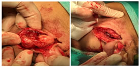
MATERIALS AND METHODS Our case history includes neoformations of the major salivary glands subjected to surgery in the period from January 1, 2008 to December 31, 2019 ( fig. 1, 2 ). Preoperative needle biopsy has not always been performed, especially in the first years of this series. In many cases in which ultrasound and magnetic resonance imaging posed for frankly benign pathology, surgical excision of the neoplasm was directly performed, entrusting the diagnosis of nature to the definitive postoperative histological examination. In the other cases, preoperative needle biopsy was performed, for a total of 40 cases. Of these 40 cases, 33 were benign neoplasms, of which 13 Warhin tumors and 20 pleiomorphic adenomas, while 7 were malignant neoformations, of which two cases of adenoid-cystic carcinoma, 4 cases of muco-epidermoid carcinoma and 1 case of acinic cell carcinoma. Figure 1, 2: Pleiomorfic adenoma of parotid gland: surgical excision OBSERVATION AND RESULTS We found in 10 cases a discrepancy between preoperative cytological examination and definitive postoperative histological examination. In two cases the preoperative finding was of pleiomorphic adenoma while the definitive one was in a case of adenoid-cystic carcinoma and in a case of mucoepidermoid carcinoma. In another 8 cases, concerning benign neoplasms, the preoperative diagnosis of Warthin tumor was changed to pleiomorphic adenoma on the definitive postoperative histological examination. The error rate was therefore 25% in total, while for malignant neoplasms, there were 2 out of 7 cases of false negatives (28.57%). Unfortunately, the FNAC does not have the same specificity and sensitivity that it presents for thyroid nodules and in pediatric age Guzzo and coll. 8,9,10 write that the needle aspiration with a thin needle is not necessary for parotid swellings since once verified with imaging methods the non-vascular nature of the neoplasm, cytology does not change the therapeutic approach that remains parotidectomy; the same author states that needle aspiration, again in children, can be "useful in submandibular and sublingual swellings to differentiate the very rare malignancies of salivary origin from other more common pathologies (lymphadenopathies etc)". and that in suspected lesions of the minor salivary glands it is better to prefer biopsy with oncotome. In the guidelines of the Italian Association of Medical Oncology (AIOM) 2008 and 2013 15 of head and neck cancers in chapter 8 (salivary gland carcinomas) it is expressly written that cytological examination with thin needle under ultrasound guidance has a modest sensitivity in detecting a malignant tumor . A second pathological evaluation is indicated, precisely because of the complexity of the diagnostic evaluation, the number of possible differential diagnoses and the paucity of the material considered in the diagnostic biopsy. For this reason, the neoplasms of this district are those in which there is a greater discrepancy between the initial pathological diagnosis and the second pathological evaluation. In the Scientific Bases for the Guidelines of the Italian Institute of Health, the chapter on Malignant Epithelial Malignancies of the major salivary glands by R. Silvestrini 24 is written tahat due to the remarkable cyto-architectural complexity of salivary neoplasms and the morphological similarities existing among numerous histotypes, FNA is reliable for diagnosing no more than 80% of salivary carcinomas . In the chapter "Tumors of the major and minor salivary glands" of the State of the Art of Oncology in Europe website (www.startoncology.net) Marco Guzzo and Laura Locati (National Cancer Institute - Milan, Italy) and Franz Josef Prott (St Josefs Hospital - Wiesbaden, Germany) state that the role of needle aspiration is important above all to avoid surgery in about a third of cases, that morbidity is very low and that cases of neoplastic implantation have not been reported but that "despite these observations the use of the aspirated needle is at the discretion of the clinician ". The reasons for these diagnostic difficulties stem from the number and complexity of the histotypes and the rarity of some of them. Marco Piemonte 19 states that morphologically similar and partly overlapping neoplastic areas are constantly observed between tumors of different histotypes, so the correct diagnostic definition of some tumors cannot derive exclusively from the examination of a few cellular aggregates, but requires the overall examination of the surgical piece or at least a significant part of it . Almost all the authors agree in avoiding the biopsy in the open air and also that with a non-thin needle due to the risk of dissemination which in the salivary glands is very high even in the case of pleomorphic adenoma. Prof Spiro 25 of the Memorial Sloan Kettering Cancer Center in New York writes that aspiration biopsy can add useful information, buti s not essential for treatment planning as the extent of the surgical procedure is primarily determined by the extent of the tumor. Prof Batsakis 2,3,17 says that while needle aspiration cytology is particularly applicable to head and neck injuries, it is not as effective in the case of the major salivary glands. Prof Theodor Miller, Director of Cytology at the University of San Francisco, California, 18 writes that ability to specifically designate what type of benign or malignant neoplasmi is present, howeer, is somewhat more problematic ... With all the difficulties in classifying salivary gland neoplasms histologically, it is easy to imagine how difficult it is to be absolutely correct on a cytologic sample For these reasons the FNAB diagnosis of salivary gland lesions can be fraught with problems and pitfails.
CONCLUSIONS We now use preoperative cytological diagnosis by means of needle biopsy in neoplasms of the major salivary glands almost constantly. Probably the margin of error of 25% or higher is destined to improve over the years both for the acquisition of better manual skills during the ultrasound-guided sampling and for the improvement of cytological techniques. However, the considerable variability of cytopathological pictures found in the context of salivary neoplasms and the frequent presence of borderline forms makes it difficult to define a precise preoperative cytological definition so we consider the data obtained by this method always with the benefit of the doubt.
REFERENCES
Policy for Articles with Open Access
|
|
 This work is licensed under a Creative Commons Attribution-NonCommercial 4.0 International License.