 Home
Home
|
Table of Content Volume 14 Issue 1 - April 2020
High sensitivity C-reactive protein vs quantitative Troponin-I as a prognostic marker in acute myocardial infarction
Anand Gaurav1, Kaushal Kishore2*
1Junior Resident, 2Professor, Department of Medicine, Patna Medical College and Hospital, Bihar, INDIA. Email: dranand21@gmail.com
Abstract Background: Acute myocardial infarction (AMI) is a major public health problem among the non communicable diseases in the developing countries like India, inspite of progressive research in diagnosis and management over the last three decades. Inflammation plays an important role in the AMI. An acute phase response C-reactive protein (CRP), is a marker of inflammatory activity and is associated with atherosclerosis and increased incidence of coronary events. Cardiac troponins are regulatory proteins within the myocardium that are released into the circulation when damage to the myocytes has occurred. Cardiac troponins has also been proven to be a potent, independent indicator of recurrent ischemic events, and an estimate for the risk of death among patients presenting with ACS. Methodology: It was observational study, it was performed at Patna Medical College, Patna, Bihar. All patients were collected from the department of Medicine IPD and to be tested in the department of central pathology based on clinical diagnosis. 60 Patients of Acute myocardial infarction, and the age group of 20-70 years was taken. Total study duration was 12 months from June 2018 to May 2019. Results: Majority of patients i.e. 66.6% patients belonged to 51-60 years in age group, ST segment elevation and Q wave myocardial infarction and Non ST segment elevation and Non Q wave myocardial infarction 58.3% and 47.1% in AMI patients found respectively. we have found significant correlation between LVIDs, EF and hsCRP. Conclusion: Both biomarkers is very important diagnostic tool of in acute myocardial infarction detection. hsCRP was positive correlation with IVS and Trop-I. hsCRP have more sensitivity than Trop –I, Trop-I have more specification compare to hsCRP. Key Word: Troponin-I
INTRODUCTION Role of hs-CRP in Acute Myocardial Infarction: Atherosclerosis has an inflammatory nature1 and results in thrombotic complications.2 Inflammatory markers, such as high-sensitivity C-reactive protein (hsCRP), are useful prognostic factors for cardiovascular events in several situations, independent of the traditional risk factors described in the Framingham risk score.3 The guidelines established by the Center for Disease Control and Prevention and the American Heart Association (AHA) in 2003 included hsCRP as a useful tool for therapeutic decision-making in selected cases.4 Coronary artery disease (CAD) is the major cause of death nearly all over the world. Atherosclerosis, which is the underlying cause of most CAD, starts early in life, progresses slowly and remains subclinical for decades. Atherosclerotic process involves the interplay of inflammatory stimuli, endothelial injury and dysfunction, enhanced adhesion molecule gene expression, monocyte attraction and activation, and secretion of multiple cytokines 5. Inflammation contributes to different stages in the pathogenesis of CAD. It plays a pivotal role in initiation, progression, and destabilization of atheromatous lesions, which eventually cause ischemic necrosis in acute myocardial infarction (MI). Several inflammatory parameters, such as cell adhesion molecules, cytokines, chemokines, and acute phase reactants, have been identified as valuable risk markers for the prediction of cardiovascular events. Among these, high sensitivity C-reactive protein (hs-CRP) has been central to most studies in the field. Hs-CRP predicts future coronary events merely because it is associated with all of the major risk factors for atherosclerosis, namely dyslipidemia, smoking, hypertension, diabetes, abdominal obesity, depression, other psychosocial factors and many others.6 Hs-CRP plays a dual role both as a sensitive, albeit nonspecific, marker of inflammation and as an active player, increasing the risk for atherosclerosis. Its plasma concentration can rise by over 1000 fold in 24-48 hours after a strong acute stimulus such as sepsis or acute MI and can fall with a half time of about 24 h when the stimulus is removed. The main source of plasma CRP is liver. Its production appears to be regulated, during the acute phase response, by several cytokines, mainly interleukin-6, interleukin-1 and tumor necrosis factor-alpha. CRP is frequently measured in clinical practice and has been investigated extensively in the context of atherosclerosis and vascular complications 6. CRP has been shown to be a prognostic marker of adverse clinical cardiac events, such as death, acute myocardial infarction, and urgent revascularization. CRP measured on hospital admission for MI was associated with a strong, positive graded increase in the risk of heart failure and death independently of known prognostic factors 7. Role of Trop-I in Acute Myocardial Infarction: Cardiac troponins have not only diagnostic value, but yield prognostic information as well. Patients presenting with clinical evidence of ischemia and increased troponins have worse outcomes than those without detectable troponin in the circulation.8 Even in patients with stable coronary artery disease, high-sensitivity assays have demonstrated that detectable concentrations of cardiac troponin portend a higher incidence of heart failure and cardiovascular death.[9] Prognosis is related in part to the extent of the increase in troponin in patients with an ischemic mechanism for myocardial injury. Recently, the MISSION! trial showed that peak troponin T levels are a good estimate of infarct size, and an independent predictor for left ventricular function at 3 months, and major adverse cardiac events at 1 year.10 Cardiac troponin has also been proven to be a potent, independent indicator of recurrent ischemic events, and an estimate for the risk of death among patients presenting with ACS. The mortality risk appears to correlate with the level of troponin rise. The TIMI-IIIB trial demonstrated that in patients presenting with ACS, mortality was consistently higher among patients with elevated troponin I at the time of admission. There were statistically significant increases in mortality with increasing levels of troponin I, with a relative risk for death of 7.8 among the group with the highest level of troponin, even after adjustment for age > 65, ST depression on ECG, and other baseline variables. Additionally, the GUSTO IIa trial found that elevated troponin T was significantly predictive of 30-day mortality in patients with acute myocardial ischemia, even after analysis was adjusted for electrocardiographic category and CK-MB level.11 Acute myocardial infarction (MI) triggers an acute phase response which is induced by proinflammatory cytokines, which are released from the necrotic myocardium and they stimulate the liver to synthesize C-reactive protein (CRP). And Myocardial infarction (MI) is defined by the presence of myocardial necrosis in combination with clinical evidence of myocardial ischemia. Cardiac troponins are regulatory proteins within the myocardium that are released into the circulation when damage to the myocyte has occurred. So our proposal, in respect to find out the importance of the prognostic marker which investigation among Hs-CRP and Trop-I if more relevant.
METHODS Type of Study: Observational study. Study Area: This study was performed at Patna Medical College, Patna, Bihar. All patients were collected from the department of Medicine IPD and to be tested in the department of Central pathology, Patna Medical College- based on clinical diagnosis. Study Population: At least 60 Patients of Acute myocardial infraction, and the age group of 20-70 years was taken from the department of Medicine IPD of Patna Medical College, Patna. All patients will be undergoing different examinations that include:
Period of Study: Total study duration was 12 months from June 2018 to May 2019. Selection Criteria: Inclusion Criteria
Exclusion Criteria:
Case Definitions
Analytical details and assays: Venous blood samples were tested for lipid profile including total cholesterol (TC), triglycerides (TG), low density lipoprotein (LDL), high density lipoprotein (HDL), and hsCRP. TG, LDL ,and HDL were analyzed by a fully autoanalyzer(Selectra pro XL) . hsCRP assays were performed with the turbidimetric assay method (Quantex hsCRP kits) The kit detected hsCRP in the range of 0.10–20.0 mg/L, all the cases of Trop-I was done outside of the laboratory. Blood samples were taken for analysis of hsCRP on admission to hospital at first AMI in the acute phase (<12 hours from onset of symptoms) from the antecubital vein. After centrifugation, the serum was separated and assayed. Statistical analysis We used the Statistical Package for Social Sciences (SPSS version 20; IBM). The demographic data and lipid profiles were expressed as the mean±SD for continuous variables and as percentage values for categorical variables. Categorical variables were compared between various groups using the correlation and coefficient . p<0.05 was considered statistically significant. RESULTS Table 1: Age and Sex Distribution (n= 60)
Age and sex distribution of AMI patients is mentioned in Table 1. We divided total patients in 5 age groups i.e. 20-30, 31-40, 41-50, 51-60 and 61-70. Majority of patients i.e. 66.6% patients belonged to 51-60 years in age group, among them 30(50%) were male and 10(16.7%) patient was female.
Table 2: Clinical Findings (n=60)
Proportion of patients with Chest pain (90%) was significantly higher among others clinical findings. We have found another clinical finding is Sweating, Shortness of breath, palpitation, vomiting and syncope i.e 70%, 63%,35%,28% and 15% respectively. Table 3: ECG Findings (n=60)
We have found in ECG-equal number of ST segment elevation and Q wave myocardial infarction and Non ST segment elevation and Non Q wave myocardial infarction 58.3% and 47.1% in AMI patients respectively.
Table 4: Mean and SD value of Lipid profile and Random Blood glucose among study population (n=60)
In table no 4, we have found the Mean and SD value of T. Cholesterol is 242.833±38.91, Triglyceride is 228.716±46.29, HDL is 25.633±±2.10, LDL is 171.456 ±37.33, VLDL is 45.743±9.25 and random blood glucose is 129.983±19.31 respectively. Table 5: Mean and SD Value of Echocardiographic Parameters study population (n=60)
Mean and SD value of different Echocardiographic parameters among MI patients is shown in Table 5.
Table 6: Pearson correlation analysis of Troponin- I and CRP levels at AMI with Echocardiography in all AMI patients
Pearson correlation analysis of Trop-I with Echocardiographic parameters in all MI patients have found, positive correlation with hsCRP, LVIDd, LVIDs, LVPWD, and LA, Negative correlation with EF. And we have found statistical correlation between LVIDs , EF and hsCRP. When correlation with hsCRP and Echocardiographic parameters , we have found positive correlation with, LVIDd, LVIDs, LVPWD, and LA, accept EF(Negative correlation). hsCRP have positive statistical correlation with IVS and Trop-I.
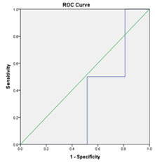
Figure: 1. Sensitivity and Specificity of hs CRP and Trop-I ROC curve analysis to determine the cutoff points and Trop-I and hsCRP levels on admission with myocardial infarction for heart failure prediction.
DISCUSSION This study reports that higher levels of hsCRP and Trop-I at admission in AMI patients predicts heart failure. Inflammatory markers are important indicators for discrimination of different clinical forms of AMI. The incidence of AMI patients was found to be highest in the age group of 51-60 yrs i.e 66.7%, We have found mean and SD value of age was 55.650±6.84. Age and sex distribution of AMI patients is mentioned in Table 2. We divided total patients in 5 age groups i.e. 20-30, 31-40, 41-50, 51-60 and 61-70. Majority of patients i.e. 66.6% patients belonged to 51-60 years in age group, among them 30(50%) were male and 10(16.7%) patient was female. Study of Gurunani RH et al. conducted12, 100 patients, 68 were male and 32 were female as shown in. The age range of this study was 30-100 years. We divided these 100 patients in 7 age groups of 10 years range. The data obtained for individual patients were arranged according to the age group of patient. In our study, proportion of patients with Chest paint (90%) was significantly higher among others clinical findings. We have found another clinical finding is Sweating, Shortness of breath, palpitation, vomiting and syncope i.e 70%, 63%,35%,28% and 15% respectively. In our study, among the patients with AMI, past history of DM (80.0%), Past history of hypertension present was (66.7 %), F/H of Ischemic Heart Disease (50%), Chronic smoker were (55%) and Alcohol abuse were (36.7%) found respectively. Clinical assessment sciences institute in Canada carried out a study on 4403 patients in Ontario Canada. They all had heart attack records which showed that mean of their age (67.3 years) and 33.7% were women. Statistics showed that numbers of married men who have been taken to hospital after heart attack faster were more than single ones, but in contrast there has been no relationship between marital statuses of women with arrival speed to hospital after experiencing chest pain related to heart attack which indicates that women take care of their husbands better (Chow et al.., 2007)13; “Iranian Students’ News Agency - ISNA,” 1998-2013). 48.4% of participants were illiterate in the present study. Results of a study showed that the majority of patients 79.1% did not have any information about the initial symptoms of MI (Akbari et al.., 2009)14. Illiteracy lead to ignorance and uneducated people certainly have lower level of health behaviors and preventive behaviors. Providing information by mass media and correct planning by health centers is needed for secondary prevention. It is recommended to provide necessary information for public education and also training and counseling for older patients with higher risk of heart disease. We have found in ECG-equal number of ST segment elevation and Q wave myocardial infarction and Non ST segment elevation and Non Q wave myocardial infarction was found 58.3% and 47.1% in AMI patients respectively. The ECG is an important initial diagnostic tool in the evaluation and management of patients presenting with chest pain. ECG findings form the basis on which acute coronary syndromes are classified into STEMI, NSTEMI, or unstable angina, a classification which provides information regarding the extent of myocardium at risk and guides initial therapy. ST-segment elevation is a sign of transmural ischemia and identifies patients who are likely to benefit from urgent revascularization. The relation between ST-segment elevation on the ECG and the occluded coronary artery has been established in multiple clinical studies in patients with ACS15. LAD coronary artery obstruction most often results in ST-segment elevation in the precordial leads V1-V4 (anterior, anteroseptal patterns)16. In rare instances, ST-segment elevation in leads V1-V4 signifies RCA occlusion with concomitant right ventricular infarction15. Isolated ST elevation in leads V4–V6, without ST elevation in leads V1–V3, is usually due to an occlusion of the LCX or distal diagonal branch rather than the main LAD artery[15]. ST-segment elevation in leads II, III, and aVF is associated with infarction of the inferior wall[15,16]. The culprit vessel in inferior MI may be either the RCA (in the majority of cases) or the LCX artery[16]. Consistent with prior studies, we found that all of our patients with anterior STEMI had LAD obstructive CAD, while among patients with inferior STEMI almost 80% had an occluded RCA and all but 1 of the remainder had an occluded LCX artery. we have found the Mean and SD value of T. Cholesterol is 242.833±38.91, Triglyceride is 228.716±46.29, HDL is 25.633±±2.10, LDL is 171.456 ±37.33, VLDL is 45.743±9.25 and random blood glucose is 129.983±19.31 respectively . Gorecki et al.17 observed higher levels of TC and LDL in patients with complicated versus those with uncomplicated clinical course of infarction, suggesting higher levels of these biomarkers during the first 24 hours of AMI have a strong negative prognostic value. Akosah et al.18 reported acceptable or optimal LDL levels in a series of 183 young adults experiencing AMI. High serum levels of HDL are associated with reduced risk for the development of atherosclerotic disease. HDL particles are believed to be antiatherogenic, secondary to their capacity to drive reverse cholesterol transport and antagonize pathways of inflammation, thrombosis, and oxidation. The majority of patients in both the primary and secondary prevention settings continue to experience significant residual risk for acute cardiovascular events, even when their LDL cholesterol is lowered aggressively via a combination of lifestyle modification and pharmacologic intervention.19 Al Aqeel et al.[20] have observed that HDL appears to be the main lipid risk factor in patients presenting with AMI in Kuwaiti patients, suggesting that primary prevention strategies should focus on treatment modalities that increase HDL. Many conditions specific to the heart in diabetes affect global myocardial remodeling. Previous silent infarction may be present in as many as 40% of these patients at the time they present with their first clinically recognized MI. Cardiac autonomic neuropathy may be present in nearly 50% of the diabetic population with coronary artery disease21 Echocardiography is an accurate noninvasive test that enables detection of evidence of myocardial dysfunction caused by ischemia or necrosis [22]. Evaluation of wall motion while a patient is experiencing chest pain can be useful when the ECG is nondiagnostic[129]. Evaluation of wall motion may also be useful if there is ECG or laboratory evidence of MI even in the absence of chest pain[23]. Severe ischemia produces regional wall motion abnormalities (RWMAs) that can be visualized echocardiographically within seconds of coronary artery occlusion (12±5 and 19±8 seconds in two series of patients evaluated during transient coronary occlusions induced by angioplasty)23. These changes occur prior to the onset of ECG changes or the development of symptoms23. The RWMAs reflect a localized decrease in the amplitude and rate of myocardial excursion, as well as a blunted degree of myocardial thickening and local remodeling. Pearson correlation analysis of Trop-I with Echocardiographic parameters in all MI patients have found, positive correlation with hsCRP, LVIDd, LVIDs, LVPWD, and LA, Negative correlation with EF. And we have found significant correlation between LVIDs , EF and hsCRP. When correlation with hsCRP and Echocardiographic parameters , we have found positive correlation with, LVIDd, LVIDs, LVPWD, and LA, accept EF(Negative correlation). hsCRP have positive significant correlation with IVS and Trop-I. The study by Kavsak et al.24 indicated that high CRP titers, independent of the subjects’ age, gender, and cTnI concentrations, predict long-term heart failure and mortality. Not only did CRP≥15 mg/L identify patients with heart failure at entry, it also predicted worsening of left ventricular function in patients presenting without clinical signs of heart failure at entry. Furthermore, in agreement with their results, CRP levels were a significant predictor of heart failure in our study in patients who did not have had any signs of heart failure at their first myocardial infarction. Although the exact role of CRP requires further exploration, our study focusing on hsCRP measurements within the first 3 days after AMI may prove useful for identification and prediction in patients who are at greater risk of heart failure and mortality. Scirica et al. have reported that both hsCRP and BNP measured 30 days after ACS are independently associated with the risk of heart failure and cardiovascular mortality, with the greatest risk occurring when both markers are simultaneously high.30 CRP has been a marker of interest as a predictor of heart failure and pharmacological treatment benefits. ROC curve analysis to determine the cutoff points and Trop-I and hsCRP levels on admission with myocardial infarction for heart failure prediction. Compared with hsCRP measurement, troponin-I assays are substantially more sensitive in the detection of myocardial injury. The release kinetics of troponins T and I are similar; both are detectable in the serum within 4 to 12 h after the onset of myocardial necrosis and depending on the duration of ischemia and reperfusion status, peak values occur 12 to 48 h from symptom onset. Therefore, serial sampling, including a baseline sample and follow-up testing at 8 to 12 h after symptom onset, is recommended. allowing for diagnostic confirmation even when patients delay presentation after symptom onset. Because of their long serum half-lives, however, neither troponin I nor troponin T assays are ideal for detection of re-infarction after an index event.
CONCLUSION hsCRP levels on admission to hospital after first myocardial infarction are associated with an increased risk of heart failure. Our study shows that inflammatory processes plays an independent role in the development of heart failure after myocardial infarction. hsCRP and Trop I are a useful markers for predicting the time course of heart failure in patients with AMI. Thus, hsCRP measurements may assist in risk stratification after myocardial infarction. Measurement of hsCRP levels in patients earlier in AMI could help clinicians to discriminate those patients who are at increased risk of heart failure in the future. Measurement of cardiac troponins is rapidly assuming the position of gold standard for the biochemical diagnosis of myocardial necrosis. At the end of the study we can conclude that both biomarkers are very important diagnostic tool of acute in myocardial infarction detection, but we have found hsCRP is more sensitive then TROP-I and the specification is more in Trop-I compeared to hsCRP.
REFERENCES
Policy for Articles with Open Access: Authors who publish with MedPulse International Journal of Community Medicine (Print ISSN: 2579-0862) (Online ISSN: 2636-4743) agree to the following terms: Authors retain copyright and grant the journal right of first publication with the work simultaneously licensed under a Creative Commons Attribution License that allows others to share the work with an acknowledgement of the work's authorship and initial publication in this journal. Authors are permitted and encouraged to post links to their work online (e.g., in institutional repositories or on their website) prior to and during the submission process, as it can lead to productive exchanges, as well as earlier and greater citation of published work.
|
|
||||||||||||||||||||||||||||||||||||||||||||||||||||||||||||||||||||||||||||||||||||||||||||||||||||||||||||||||||||||||||||||||||||||||||||||||||||||
 This work is licensed under a Creative Commons Attribution-NonCommercial 4.0 International License.