 Home
Home
Official Journals By StatPerson Publication
|
Table of Content - Volume 11 Issue 2 - August 2019
. Safety and efficacy of intracameral mydriatics in cataract surgery in Salem district
B Jayaprakash1, R Rajeshkannan2*, K Ezhilvendhan3
1Assistant Professor, 2Associate Professor, 3Professor and HOD, Department of Ophthalmology, Vinayaka Mission’s Kirupanda Variyar Medical College and Hospital, Vinayaka Missions Research Foundation, Deemed To Be University, Salem Tamil Nadu, INDIA Email: doctorrajeshkannan@gmail.com
Abstract Background: In order to perform cataract surgery, adequate dilatation of the pupil is essential. This is traditionally achieved by preoperative topical mydriatic eye-drops, commonly cyclopentolate and phenylephrine. This routine has several disadvantages. First, the slow penetration through the cornea delays the onset of mydriasis. Second, the limited bioavailability of topically administered substances with significant systemic absorption may increase the risk for systemic side effects. Aims of the study: To evaluate an alternative mydriatic regimen for phacoemulsification cataract surgery: intracameral injection of mydriatics mixed with lidocaine (ICM). Additionally, to determine the correlation between early transient postoperative corneal edema and permanent ECL after phacoemulsification cataract surgery. Methods: The study was conducted prospectively in Department of Ophthalmology in Vinayaka mission’s Kirupanda Variyar medical college and hospital, vinyaka missions research foundations deemed to be university, Salem, Tamil Nadu for a period of six months Sixty patients were included and randomized. One group (30 patients) was given TM comprising 3 drops each of cyclopentolate 1% and phenylephrine 10% at 15-minute intervals before surgery. They also received 150 µl of preservative-free lidocaine 1% intracamerally at the beginning of the procedure. The other group (30 patients) was given placebo eye drops at the same intervals as in the topical group and ICM at the start of the operation. Pupil measurements were made every 5 seconds after the ICM injection. The pupil diameter was also measured after OVD injection before capsulorhexis and before and after OVD injection prior to the IOL insertion. Results: With ICM, mydriasis reached 95 ± 3% of its final value within 20 seconds. In the ICM-group, the pupils were smaller than in the TM-group (mean 6.7 ± 1.0 mm versus 7.7 ±1.0 mm, P<.001), but did not contract intraoperatively as the TM pupils did. Conversely, with ICM the pupil sizes generally increased during the cataract procedures. This increase was significantly greater without epinephrine in the irrigating solution (13 ± 19% versus 4 ± 14%; p = 0.02). No significant differences in pupil sizes were observed between the patients who were given ICM with or without cyclopentolate. The central corneal swelling at the first postoperative day was strongly correlated to the central ECL at 3 months, R2 = 0.785, P < 0.001. Conclusions: ICM is a rapid and safe alternative to TM in phacoemulsification cataract surgery. An irrigating solution without epinephrine can safely be used with ICM. Cyclopentolate administrated intracamerally, has no immediate additive mydriatic effect to intracameral lidocaine combined with phenylephrine. The degree of permanent corneal endothelial damage in cataract surgery is reflected in the degree of early postoperative corneal swelling. Key Word: Intracameral, Mydriatics, Cataract, Phacoemulsification, Cyclopentolate - Epinephrine, Endothelial Cell Loss.
The cornea, the iris, and the lens are situated in the anterior part of the eye. The main function of the cornea is to refract the incoming light and it contributes to approximately 2/3 of the total refractive power (the lens refracts the remaining part).1 The cornea is made up of three major structures. The epithelium consists of 4-6 cell layers that form 5-10 % of the total corneal thickness of about 540µm. The superficial epithelial cells have tight junctions that prevent penetration of tear fluid into the stroma. The stroma mainly consists of collagen lamellae with interleaved keratocytes and constitutes about 90% of the corneal thickness.2 Its water content differs in different parts of the structure and the transparency is dependent on its dehydrated state, which in turn is maintained by the endothelial pump. The endothelium is a 5 µm thick monolayer of closely joined hexagonal cells.3 From what we know today, these cells are not likely to proliferate in humans and the loss of cells is therefore compensated by sliding and thinning of adjacent cells to cover the defect. This lack of cell division results in a gradual corneal endothelial cell loss (ECL) throughout life, with an average cell loss of 0.3-0.6% per year. At birth, the endothelial cell density is 3500-4000 cells/mm2 and decreases in adulthood to 1500-2500 cells/ mm2. Apart from the ECL in the normal aging process a reduction in cell number can also occur due to trauma intraocular surgery 37-39 and ocular diseases.4 A cataract is any opacity of the lens, whether it is a small local opacity or a diffuse general loss of transparency. To be clinically significant, however, the cataract must cause a significant reduction in visual acuity (VA) or functional impairment. Symptoms of cataract include reduced visual acuity, photophobia, glare, motivation, and monocular diplopia. A cataract is one of the commonest causes of vision loss and the worldwide leading cause of blindness. WHO has estimated that 40-45 million people worldwide have a non-useful vision (i.e. they are unable to walk about unaided or have a VA <3/60) and that cataract accounts for 48 % of these cases. Population-based studies in the USA have shown that the prevalence of cataract, causing loss of vision to 20/30 or worse in persons 75 years or older, is about 39 % for men and 46 % for women.5 For all ages, the prevalence is 16 % and for those older than 75 years 42%.17 Without consideration of VA, the prevalence for senile lens opacities are for ages 52-64 years 21%, 65-74 years 53% and 75-85 years 80%. In 2006, approximately 72,500 cataract operations were performed in Sweden and 62% of these were carried out in women.6 MATERIALS AND METHODS The study was conducted prospectively in Department of Ophthalmology in Vinayaka mission’s Kirupanda Variyar medical college and hospital, Vinayaka missions research foundations deemed to be university, Salem, Tamil Nadu Sixty patients were included and randomized. The collected cases were classified according to gender-wise, age-wise, a number of surgeries in each month, affected the eye, visual acuity and the type of cataract affected. The parameters like blood pressure, pulse rate, and pupil size pre and intraoperatively were measured to determine the safety and efficacy of intracameral Adrenaline during phacoemulsification. In order to maintain mydriasis 1ml of Adrenaline (0.00001%) was given intracamerally after corneal incision. The solution was prepared prior to the surgery by the surgeon given intracamerally just after corneal incision and let it to stay at the anterior chamber for 1min. Pre and intraoperative parameters were measured, and the subjective surgical performance was graded after each procedure. Procedure: Sixty patients were included and randomized. One group (30 patients) was given TM comprising 3 drops each of cyclopentolate 1% and phenylephrine 10% at 15-minute intervals before surgery. They also received 150 µl of preservative-free lidocaine 1% intracamerally at the beginning of the procedure. The other group (30 patients) was given placebo eye drops at the same intervals as in the topical group and ICM at the start of the operation. Pupil measurements were made every 5 seconds after the ICM injection. The pupil diameter was also measured after OVD injection before capsulorhexis and before and after OVD injection prior to the IOL insertion. Two surgeons performed each operation so that one could start the procedure and give the intracameral injection; thus, neither the subsequent operating surgeon nor the patient was aware of which treatment was given in each case. Preoperatively and 1 day and 1 month postoperatively, the pupil size and the central, nasal, and temporal corneal thicknesses (pachymetry) were measured with the Orbs can II and central corneal endothelial photographs were taken with the Topcon SP-2000P specular microscope. Statistical Analysis: All the values were expressed as mean ± SD and P < 0.05 was considered statistically significant. Statistical significance of the differences between the mean values was analyzed by one-way ANOVA test using SPSS 16 statistical analysis software. Correlations between different variables were analyzed using Pearson’s correlation coefficients (r). Sample size was calculated using (α = 0.05, β = 0.2, σ = 18, d = 11) formula.
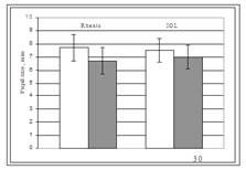
RESULTS Figure: 1 In the group which received ICM, the pupils reached 95 ± 3 % of their maximum size after 20 seconds (Figure 1). The mean pupil size after OVD injection was smaller in the ICM group (6.7 ± 1.0 mm) than in the TM group (7.7 ± 1.0 mm) (P < 0.001), but the pupils in the ICM group often continued to enlarge throughout the cataract procedure (+4.5 ± 8.1%). Conversely, in the TM group the pupils tended to contract (−2.1% ± 7.8%) (P = 0.002). The difference in pupil size before IOL implantation way, therefore, smaller (7.0 ± 0.9 mm vs.7.5 ± 0.9 mm) between the groups, but still significant (P = 0.04) (Figure 2). At 1 day, the pupils in the ICM group were significantly larger than in the topical group (5.7 ± 1.1 mm versus 3.7 ± 0.8 mm) (P < 0.001). Figure 1 Figure 2 Figure 1: Pupil Size (Mean ± Sd) After Injection Of Icm (S = Seconds), N=30; Figure 2: pupil size at capsulorhexis (rhexis) and before iol implantation (iol).white bars – tm (n=30). Grey bars – icm (n=30) Graph :2 The mean ECL at 1 month was 2.3 ± 6.1% in the ICM group and 4.1 ± 6.0% in the TM group (P = 0.25) (Table 1), but the postoperative cell counts were not significantly different from the preoperative values (P = 0.2 and P = 0.2, respectively). In contrast, cells were more irregular and elongated postoperatively in both the ICM group and the TM group as assessed by HSF (P = 0.009 and P = 0.001, respectively) and by DE (P = 0.004 and P < 0.001, respectively). Again, there were no significant differences between the two groups (Table 1).A significant decrease in pulse rate occurred in the TM group (P = 0.0055) but not in the ICM group (P = 0.15). The difference in pulse deceleration was significant between the groups (P = 0.009) Graph :2 The mean ECL at 1 month was 2.3 ± 6.1% in the ICM group and 4.1 ± 6.0% in the TM group (P = 0.25) (Table 1), but the postoperative cell counts were not significantly different from the preoperative values (P = 0.2 and P = 0.2, respectively). In contrast, cells were more irregular and elongated postoperatively in both the ICM group and the TM group as assessed by HSF (P = 0.009 and P = 0.001, respectively) and by DE (P = 0.004 and P < 0.001, respectively). Again, there were no significant differences between the two groups (Table 1).A significant decrease in pulse rate occurred in the TM group (P = 0.0055) but not in the ICM group (P = 0.15). The difference in pulse deceleration was significant between the groups (P = 0.009)
Table 1: Selected Perioperative Parameters.
DISCUSSION Peri- and postoperative parameters such as surgical time, amount of irrigation fluids, cells and flare in the anterior chamber, IOP and VA are not different compared with traditional TM. Patients in the intracameral group also reported less discomfort from glare. This is probably due to that a normal-sized pupil at the initiation of the procedure allows a more gradual adaptation to the microscope light.7 Because of the design of study I, the mydriatics and lidocaine were often left in the anterior chamber for several minutes before surgery.8 From the, we indirectly conclude that leaving ICM in the anterior chamber for a few minutes is unlikely to have detectable adverse effects on the eye. A possible advantage with ICM is that the preoperative time to prepare the patient is shortened and the flexibility, i.e. to switch orders of the patients, is improved. 9 This is helpful both for the preparing staff and the doctor and may lead to better working conditions and expanded operation schedules10 The phaco CDE was significantly lower in the ICM group study. This could mean that the lenses were softer in this group, although the grade of cataracts was similar between the groups. A lower phaco CDE and an equal operation time could also indicate that the surgical performance is as good with ICM as with TM, even though in general, the pupils were slightly smaller in the intracameral group11 Furthermore, the pupils dilated with ICM often continued to enlarge during the cataract operation. Therefore, clinically with ICM, if the pupil is sufficiently large at the beginning of the procedure, it is likely to remain sufficiently large throughout the procedure.12 A pupil that enlarges during surgery, even if it is smaller, may even be more comfortable to work with than a larger pupil that is contracting.13 The lower doses of mydriatics with ICM may, therefore, make it especially suitable in certain cases such as patients with heart conditions Adding epinephrine to the irrigating solution does not contribute to preserving the pupil size during phacoemulsification cataract surgery when using ICM. On the contrary, a greater pupil enlargement is achieved with ICM without epinephrine in the irrigating solution than with epinephrine.14 The exact mechanisms behind this finding are not known, but the dilator muscle in the iris has mainly α-adrenergic and few β-adrenergic receptors. Phenylephrine is more potent in stimulating α-receptors than epinephrine. The response occurs in reverse order for β-receptors. 15 When phenylephrine is injected into the anterior chamber, the adrenergic receptors become occupied, but when epinephrine enters the eye it may gradually compete with phenylephrine and take over some of the receptors. Being a weaker dilator, epinephrine may thus lead to a smaller pupil.16 The difference in pupil size when using ICM with or without epinephrine in the irrigating solution, become less pronounced after injection of OVD as the OVD acts as an unspecific pupil dilator.17
CONCLUSIONS The sixty patients were followed up to one month. However, it may be difficult to demonstrate a significant cell loss without a larger number of patients. On the other hand, we observed a greater cell irregularity postoperatively that may indicate that the endothelial cells had not “come to rest” completely and that a follow-up of 1 month is too short to make conclusions. ICM with lidocaine 1%, phenylephrine 1.5%, and cyclopentolate 0.1% is a new method of dilating the pupil before cataract surgery. It is a safe and effective way to achieve an adequate pupil size in order to carry out the cataract procedure.
REFERENCES
|
|

 This work is licensed under a Creative Commons Attribution-NonCommercial 4.0 International License.