 Home
Home
Official Journals By StatPerson Publication
|
Table of Content - Volume 11 Issue 2 - August 2019
Clinical profile of patients with refractive errors in pre-presbyopic age group in tertiary care centre
Vinod Dhiman1*, Rajeev Tuli2, R K Sharma3, Sonia4, Indu Dhiman5
1Medical Officer, Civil Hospital Nurpur, Himachal Pradesh, INDIA. 2Professor & HOD, 3Professor, 4,5Senior Resident, Department of Ophthalmology, Dr RPGMC, Tanda, Kangra, H.P., India. P.C.-176001 Email: vinoddhiman77@gmail.com
Abstract Background: Refractive errors constitute a major portion of visual impairment globally. Aim of this study was to study the pattern of refractive errors in pre-presbyopic patients and its association with selected variables (age, sex, educational status, occupational status, clinical symptoms, amblyopia and strabismus). Material and Methods: The present study was an institutional based cross sectional study done on patients with uncorrected refractive errors conducted in the Department of Ophthalmology Dr RPGMC Kangra at Tanda, Himachal Pradesh. Patients in the pre-presbyopic age group (5-39 years) with uncorrected or inadequately corrected refractive error who attended Ophthalmology OPD, Dr RPGMC, Kangra at Tanda in one year i.e. from June 2016 to May 2017 were included. Data thus collected was entered and analysed using SPSS version 21 software and descriptive statistics were analysed using proportions and Chi- square test. Results: Out of total 397 subjects, 178 (44.8%) were males and 219 (55.2%) were females. The mean age of male and female patients was 19.02 ± 8.64 years and 21.03 ± 8.45 years respectively. 51.1% of the patients had secondary level of education. Occupational status wise, 67.3% of total patients were students followed by housewives. Headache was the commonest presenting clinical symptom (62%) followed by defective vision for distance and other symptoms. Among all types of refractive error, astigmatism was most common refractive error(47.7%) followed by myopia(27.30%) and hypermetropia (22.40%) and no error(2.90%). 77.5% of total patients had mild refractive error (<1.5D). Conclusion: Uncorrected refractive error is a major cause of visual impairment in all ages which can be easily diagnosed, measured and corrected. Key Word: pre-presbyopic.
INTRODUCTION Uncorrected refractive error is a leading cause of moderate and severe visual impairment globally.1 It causes adverse effect on general quality of life, affects performance in school and reduces employability and productivity. It constitutes 48.99% of the total causes of global visual impairment in all ages (blind and moderate severe visual impairment), 53.72% of total causes of moderate and severe visual impairment and 20.62% of total causes of global blindness in all ages 1 Refractive errors can be easily diagnosed, measured and corrected with spectacles or other refractive corrections to attain normal vision. If, however, refractive errors remain uncorrected or the correction is inadequate, it will result in an impaired or decreased quality of life for millions of people worldwide, irrespective of their age, sex, and ethnicity.2 The new Global Action Plan (GAP) is now the most important strategic document in eye health. Global Action Plan has a global target – the reduction in prevalence of avoidable blindness and visual impairment by 25% by 2019 from the baseline of 2010.3 The World Health Organisation Prevention of Blindness and Deafness Programme has carried out a systematic search and review of all available data to obtain a global estimate of visual impairment for 2010; for age group 5 to 14 years the global prevalence was 1.02% with highest prevalence in South East Asia and China. A prevalence of 2.3% was estimated for age group 16 to 49 years and 13.9% people aged 50 and above were visually impaired.(4) In India, 5.3% of people in all age groups were visually impaired which constitutes 21.9% of the global visual impairment. The total low vision and blindness were 4.6% and 0.7% respectively which constitutes 22.2% and 20.5% of the total global estimate respectively 4. Study from urban population in southern India which was extrapolated to the urban population of India, showed that among those more than 15 years of age an estimated 49.3 million people would have refractive error and that under corrected refractive error was the most common cause of reversible blindness.5 As per the National Programme for Control of Blindness (NPCB) survey in 2001-02, the prevalence of blindness was estimated to be 1.1%. Rapid Survey on avoidable blindness conducted under NPCB during 2006-07 showed reduction in the prevalence of blindness from 1.1% (2001-02) to 0.7% (2010-11). Various activities/initiatives undertaken during the Five Year Plans under NPCB are targeted towards achieving the goal of reducing the prevalence of blindness to 0.3% by the year 2020(6). In the age group 5–15 years, non-correction of refractive errors is due to lack of screening, lack of availability and affordability of refractive corrections. However, cultural disincentives also play a role, as shown in surveys from countries where routine screening and provision of corrections are free of charge or easily accessible, but compliance still remains low.7,8 Studies have reported that even in economically advantaged societies, refractive errors can go undetected or uncorrected in children.9 MATERIALS AND METHODS The present study was an institutional based cross sectional study done on patients with uncorrected refractive errors conducted in the Department of Ophthalmology Dr RPGMC Kangra at Tanda, Himachal Pradesh. Patients in the age group 5-39 years with uncorrected or inadequately corrected refractive error who attended outpatient clinic of Ophthalmology, Dr RPGMC, Kangra at Tanda in one year i.e. from June 2016 to May 2017 were included. INCLUSION CRITERIA Patients with visual acuity was 6/9 or less on the standard Snellen’s chart and showed improvement of one or more lines in distant vision by pin hole or 6/6 with asthenopic symptoms. EXCLUSION CRITERIA Patient with diminution of vision because of any organic cause i.e. history of cataracts, pathology of media, fundus or anterior segment, history of trauma or previous eye surgery, pseudophakia, optic nerve involvement or other causes for diminution of vision. For visual acuity Snellen’s chart for distant vision and Jaeger’s chart for near vision was used. Objective refraction was done with Streak retinoscope (Heine beta 200) and Auto Refkeratometer (Accurel-K 9001).For measuring correcting lens power AutoLensmeter GL-7000 was used. The demographic information of these patients such as name, age, sex, occupation, socioeconomic status and address was taken. Visual complaints about difficulty in reading /defective vision for distance or near/ goes close to television/ unable to see blackboard/ frequent fall was recorded. Ocular complaints like heaviness/pain in eyes/recurrent redness /deviation of eyes/ watering/recurrent swelling of lids/ recurrent stye or chalazion/frequent blinking or rubbing of eyes was also noted. Statistical Analysis: Data thus collected was entered and analysed using SPSS version 21 software. Descriptive statistics were analysed using proportions and Chi- square test. Quantitative variables were analysed using mean and standard deviation. A p value less than 0.05 was taken to be statistically significant. This was an institutional based cross-sectional study on patients with uncorrected refractive errors that was conducted in the Department of Ophthalmology Dr RPGMC Kangra at Tanda, Himachal Pradesh. A total of 397 subjects were included in the study during June 2016 to May 2017.
RESULTS Data thus collected was entered and analysed using SPSS version 21 software. Descriptive statistics were analysed using proportions and Chi- square test. Patients included in the study were in age group of 5 to 39 years. Maximum patients were in age group 10- 14 years (23.9%) followed by 15- 19 years (20.9%) age group as shown in Table 1 and Figure 1. The mean age of all patients was 20.13 ± 8.59 years and the mean age of male and female was 19.02 ± 8.64 years and 21.03 ± 8.45 years respectively.
Table 1: Age distribution of patients (n=397)
Figure 1: Histogram of age distribution of patients (n=397) the mean age of male and female was 19.02 ± 8.64 years and 21.03 ± 8.45 years respectively. i.e. had secondary level of education as shown in Table 2. No patient was illiterate. Females however tended to have a higher level of education than males. Religion wise maximum patients were Hindu in the study (98.70%) Table 2: Educational status of patients (n=397)
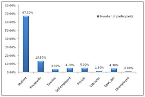
Occupational status wise, out of total patients, majority of them were students followed by housewives in the study as shown in Figure 2. Least percentage was of unemployed. Figure 2: Occupation status of patients (n=397) On basis of clinical symptoms, out of all asthenopic symptoms, headache was most common symptom followed by defective distant vision as shown in Table 3. Many subjects had more than one asthenopicsymptoms. On basis of clinical symptoms, out of all asthenopic symptoms, headache was most common symptom followed by defective distant vision as shown in Table 3. Many subjects had more than one asthenopicsymptoms.
Table 3: Distribution of various clinical symptoms of subjects
Among all types of refractive error, astigmatism was most common refractive error(Right eye 47.1% and Left eye 47.7%) followed by myopia and hypermetropia as shown in figure 3. Figure 3: Types of refractive error in both eyes of subjects (n=794) Among astigmatic refractive error in both eyes, simple myopic astigmatism was commonest followed by compound myopic astigmatism as shown in Table 4. Table 4: Types of astigmatic refractive error in both eyes of patients (n=376)
Out of 397 patients, majority of patients eyes had mild degree of refractive error as shown in Table 5
Table 5: Degree of refractive error in both eyes of patients (n=794)
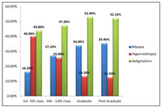
Out of total patients, myopia and astigmatism was higher in graduates and post graduates while hypermetropia was predominant in subjects with primary level of education. There was statistically significant association between education and refractive error(P value =0.001) as shown in Figure 5. Figure 4: Education wise distribution of refractive errors of both eyes of patients; Figure 5: Association between headache and types of refractive error (n=771) Figure 6: Degree of refractive errors in subjects with headache (n=771) Headache was higher in subjects with astigmatism, while in those with myopia and hypermetropia (p=0.001) as shown in 89.30% of the patients with headache had mild degree of refractive error and the rest patients had moderate to severe and very severe degree of refractive error (P=0.001). In all types of refractive errors, in majority of subjects, visual acuity was 6/18- 6/6. Hypermetropia was commonest cause for visual acuity less than 3/60 which was statistically significant as shown in table 6.
Table 6: Association between visual acuity and types of refractive error
DISCUSSION Uncorrected refractive error is one of the leading causes of moderate and severe visual impairment. It impairs the quality of life and limits the career choices, job opportunities of those affected, thus constituting a socioeconomic burden on society. However, most refractive errors can be easily corrected with appropriate spectacles or other optical devices. The present study was an institution based cross sectional study of clinical profile of the uncorrected refractive errors involving the age group of 5 years to prepresbyopic age i.e. 39 years. In this study, clinical profile of patients with uncorrected refractive errors including myopia, hypermetropia, astigmatism and their relationship with age, sex, educational status, occupational status, clinical symptoms were studied. Till date, most of the studies done to analyze the pattern of refractive errors in children were either school screening or population based and required huge economic resources. The present study being a hospital based study is unique as it has been conducted in the OPD premises without the need of extra manpower and equipments of extra manpower and equipments. In the present study, 44.8% subjects were males and 55.2% were females. There was higher proportion of females than males. In a population based study done by Dulani et al12 from Jaipur, Pavithra et al 13 from Bangalore and Prema et al 14 from Tamil Nadu showed female preponderance. Similar result was seen in hospital based study done by Tuladhar et al 15 from Nepal. It may suggest that there are no obstacles or inhibition in the health seeking behaviour between gender, and particularly that female are not prevented or side-lined in eye care provision specifically and accessibility to health care in general. In the present study, majority of the subjects were between 10 to 24 years of age (62.7%). The mean age of male subjects was 19.02 ±1.28 years and for female subjects was 21.03 ±1.13 years. Out of total subjects, maximum were students (67.3%) followed by housewives (13.4%). This finding is consistent with a study by Tuladhar et al 15 from Nepal in which majority of the subjects were students. It may be because students and younger age group sought more ophthalmologist advice for difficulty to see blackboard in the classroom. On education wise distribution of refractive error, we found that prevalence of astigmatism and myopia was more in subjects with higher level of education (graduate and post graduate) and hypermetropia in those with primary level of education. Similar results were seen in study by Dandona et al 5 from southern India. In our study, we found that headache was the commonest symptom (62.0%) in subjects followed by defective distant vision (53.4%) and pain in eyes (37.0%). A study by Gupta et al 16 from central India in children showed similar results. Prevalence of headache was more in subjects with astigmatism (51.90%) than that of myopia (22.80%) and hypermetropia (25.30%). A study by Akinci et al 17 also showed that prevalence of astigmatism was significantly higher in the headache group. In present study it was also found that headache was more prevalent in mild degree (< 1.5D) of refractive errors (87.8%). In present study, 70.8% of right eye and 71.8% of left eye had presenting visual acuity 6/18 to 6/6, 23.9% for right eye and 22.4% for left eye had presenting visual acuity 6/60 to 6/18, 4.5% of right eye and 4.3% of left eye had severe visual impairment i.e. presenting visual acuity 6/60 to 3/60 and 0.8% of right eye and 1.5% of left eye had presenting visual acuity less than 3/60. After correction of refractive error, the number of subject’s eyes with visual acuity more than 6/18 was increased to 97.5% from 71.3%. These results were similar to Malu et al 18 study. In our study, out of 794 eyes (397 subjects), astigmatism was commonest refractive error (47.4%) followed by myopia (27.3%) and hypermetropia 22.4%). Among eyes of subjects with astigmatism, simple myopic astigmatism was most common (20.5%) followed by compound myopic astigmatism (15.0%), simple hypermetropic astigmatism (6.4%), compound hypermetropic astigmatism (2.8%), and mixed astigmatism (2.7%). Our findings were in concurrence with studies by Srivastava et al 19 and Rizyal et al 20. Studies done by Dandona et al5 Qureshi et al 21 and Tuladhar et al 15 showed that myopia was most common refractive error. It may be because being a tertiary level hospital; most of the patients who came here were either inadequately corrected or referred from primary or secondary care centres. Therefore, myopic or hypermetropic eyes were less in number as compared to eyes with astigmatism, as that can easily be corrected at peripheral hospitals. In 5 to 14 years age group, we found hypermetropia was highest followed by astigmatism and myopia and in 10 to 24 years age group myopia was highest, followed by astigmatism and hypermetropia. However above 25 years, astigmatism was significantly higher. There was significantly higher hypermetropia in more than 30 years of age as compared to 20 to 29 years of age group. This result was consistent with study by Pavithra et al.13 The increase in hypermetropia beyond 30 years of age seen in our population could be explained by decrease in residual accommodation, oradecrease in the power of the aging lens, or an increase in the density of the cortex that makes the lens more uniformly refractive. These findings were in agreement with previous study by Dandona et al 5 from India. In current study, most of patients suffered from mild to moderate degree of refractive error. Myopia up to 2.75 D and hypermetropia and astigmatism up to 1.5 D were present in majority of the eyes. These findings were in agreement with studies by Hashemi et al 22, Krishnamurthy et al 23 and Shrestha et al 24. In our study, it was also found that more number of hypermetropic eyes had severe degree of refractive error.
CONCLUSION Uncorrected refractive error is a major cause of visual impairment in all ages which can be easily diagnosed, measured and corrected. We found more number of patients with astigmatic error in our study which can be corrected by early diagnosis and prescribing corrective spectacles. By this we can reduce the burden of visual impairment amongst patients who utilize eye care services. In our study, refractive error was more common in young patients. We also found headache was most common asthenopic symptom in mild type of astigmatic error. By doing complete work up including refraction in cases of headache especially among young patients, we can detect and correct refractive errors.
REFERENCES
|
|
||||||||||||||||||||||||||||||||||||||||||||||||||||||||||||||||||||||||||||||||||||||||||||||||||||||||||||||||||||||||||||||||||||||||||||||||||||||||||||||||||||||||||||||||||||||||||




 This work is licensed under a Creative Commons Attribution-NonCommercial 4.0 International License.