 Home
Home
Official Journals By StatPerson Publication
|
Table of Content - Volume 11 Issue 2 - August 2019
Optic nerve head drusen mimicking optic disc oedema – A case report
Vandna Sharma1*, Rajeev Tuli2, Gaurav Sharma3, Mandeep Tomar4
1,2,3,4Department of Ophthalmology, Dr RPGMC, Tanda, Kangra, H.P, INDIA. Email: drrajeevtuli@gmail.com, drgaurav.sharma@gmail.com, sunnyigmc85@gmail.com, vandna.27@rediffmail.com
Abstract Optic nerve head (ONH) drusen are abnormal globular acellular collections which typically occur in small, crowded optic discs with abnormal vasculature1. They may appear on surface or buried within the disc2. On Ophthalmoscopy, superficial drusen are visible as yellow highly reflectile, beige, rounded deposits. However, when they are buried within the ONH, discs may appear elevated with indistinct, irregular disc margins, mimicking optic disc edema1,4. So, it can be challenging to differentiate false edema from true swelling of the ONH, more so in a absence of other clinical signs/symptoms of ONH pathology. But, newer ocular imaging techniques are proved to be helpful in differentiating these two conditions of ONH. By doing so, unnecessary patient costs and anxiety in the case of pseudopapilledema can be reduced, and appropriate urgent referral and management in the case of true swelling of the ONH can be initiated4,5. We present here a case of bilateral ONH drusen who was referred as a case of chronic headache and to rule out papilledema as cause.
INTRODUCTION Optic nerve head (ONH) drusen are abnormal globular acellular collections which typically occur in small, crowded optic discs with abnormal vasculature1. They may appear on surface or buried within the disc2. On Ophthalmoscopy, superficial drusen are visible as yellow highly reflectile, beige, rounded deposits. However, when they are buried within the ONH, discs may appear elevated with indistinct, irregular disc margins, mimicking optic disc edema1,4. So, it can be challenging to differentiate false edema from true swelling of the ONH, more so in a absence of other clinical signs/symptoms of ONH pathology. But, newer ocular imaging techniques are proved to be helpful in differentiating these two conditions of ONH. By doing so, unnecessary patient costs and anxiety in the case of pseudopapilledema can be reduced, and appropriate urgent referral and management in the case of true swelling of the ONH can be initiated4,5. We present here a case of bilateral ONH drusen who was referred as a case of chronic headache and to rule out papilledema as cause.
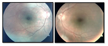
CASE REPORT A 40- year, old female initially presented to her local physician complaining of headache. She was referred to us as a suspected case of optic disc edema for further evaluation. She was also complaining of diminution of vision B/E both for near and distant. Her visual acuity was in R/E 6/9 and L/E 6/12 improving to 6/6 with pin hole in B/E. Her retinoscopy was done and correction for near was given. Her dilated fundus examination revealed small, hyperaemic, elevated discs with "lumpy-bumpy" appearance, blurred margin, absent optic cup, with no surrounding retinal oedema or any vascular abnormalities. A suspicion of B/L optic disc drusen was kept (Figure1). Medullated nerve fibres were present along the superior vascular arcade in R/E and along inferior vascular arcade in L/E. Her B-scan ultrasonography was done revealed highly reflective round structures in both eyes suggestive of disc drusen (Figure2). To further confirm the diagnosis spectral domain optical coherence tomography (SD-OCT) was done, which shows elevation of disc with irregular internal contour of optic nerve head and abrupt decline in the subretinal hypo reflective space (SHYPS) thickness. Retinal nerve fibre layer (RNFL) was of normal thickness with absent “lazy V” pattern which ruled out presence of optic disc edema (Figure 3).
Figure 1: Shows fundus photograph
Figure 2: shows B-scan shows highly reflective round structures Figure 3: shows SD-OCT images optic nerve head showing “lumpy-bumpy” internal contour of the optic nerve head and abrupt decline in SHYPS thickness suggestive of optic nerve head Drusen.
DISCUSSION Optic Nerve Head Drusen (ONH) are often incidental findings discovered during routine eye examinations. Drusen are depositions of mucopolysaccharides and proteinaceous material that accumulate anterior to the lamina cribosa within the optic nerve head which is believed to be the depositions as a result of axonal degeneration of the optic nerve6,7 or axoplasmic transport stasis secondary to congenitally crowded optic nerve heads8. ONHD are found in approximately 1% of the general population, with bilateral distribution in 75-85%2,9. They are more common among Caucasians 10. However, there pattern of inheritance remains unproven and some studies suggest the condition is transmitted as an "irregularly dominant fashion"11. The primary risk factor for developing ONHD might be inherited ONH dysplasia and its blood supply which predispose to formation of optic disc drusen2, 12. There are two main types of ONHD: visible drusen (as in this case) and "buried" drusen which are often found in younger age children and can obscure the edges of the optic disc and cup resulting in the appearance of papilledema, which is a major part of the differential diagnosis 10.With the age, drusen become more visible and begin to protrude from the edge of the optic disc and cup, especially on the inferior nasal side 6. It can compress and compromise nerve fibres and the vascular supply, leading to multiple complications including visual field defects, vascular occlusion, and haemorrhage 9. The earliest symptoms of ONHD are visual fields defects are found most commonly inferior nasal area and enlargement of the blind spot with or without constriction of the peripheral fields or transient visual obscuration, such as flickering or "graying out" 2,7,10. Additionally, many studies suggest that the presence of ONHD makes the nerve more susceptible to glaucomatous damage at lower pressures. So, one must carefully follow visual fields in suspicious cases and treat elevated IOP appropriately13. Vascular compromise to the ONH may result in Anterior Ischemic Optic Neuropathy (AION)10. Visual inspection alone is often insufficient to confirm ONHD and rule out more concerning possible diagnoses. So, it is mandatory that the physician distinguish pseudopapilledema from true papilledema. In some cases this will require appropriate radiologic neuroimaging to rule out intracranial mass or lesion.13 Diagnostic ocular imaging techniques for evaluation of ONHD are : 1) B-scan echography- is very reliable due to the characteristic highly reflective nature of drusen 10. 2) CT scans- are costly and slices of 1.5mm resolution are likely to miss the drusen. However, calcified drusen may be noticed13. 3) Fluorescein Angiography – drusen are highly auto-fluorescent and show well-defined, uneven hyper-fluorescence, especially in the late phase. This can be helpful in differentiating optic disc drusen from true papilledema, with the former showing only scarce teleangiectatic vessels2. 4) SD-OCT – useful in detecting early nerve fibre thinning and quantify any degree of nerve fibre loss with satisfying reproducibility2. Patients with documented ONHD should be followed with serial visual fields, optic nerve fibre analysis, and repeat IOP checks, no urgent medical attention is commonly required 2,10. In contrast, true swelling of the ONH secondary to increased intracranial pressure, are potentially sight and life threatening, demands accurate diagnosis and urgent management5.
CONCLUSION The ONH elevation results either from benign elevation due to ONH drusen or from true optic disc edema, such as in papilledema due to raised intracranial pressure. Both these conditions have overlapping features and hence it is imperative that these are accurately differentiated in clinical practice, as misdiagnosis has significant implications for patient management15. If clear signs and symptoms associated with optic disc oedema, medical history and a thorough eye examination are not sufficient to point to a definitive diagnosis, then different ocular imaging techniques are adjunctive which can be useful to assess precisely the disc features4. No definitive treatment currently exists for optic nerve drusen except routine monitoring where as true papilledema require urgent intervention. Hence, correct diagnosis of pseudopapilledema is important to avoid medical costs on unnecessary neurologic procedures and prevent any needless patient anxiety5.
REFERENCES
|
|


 This work is licensed under a Creative Commons Attribution-NonCommercial 4.0 International License.