 Home
Home
Official Journals By StatPerson Publication
|
Table of Content - Volume 12 Issue 1 - October 2019
Intraoperative cause of aphakia
M Samuel Christopher1, R Sujani Sunkesula2, Y Jaya Santhi latha3*
2Senior Resident ,3Professor, Department of Ophthalmology: Nizamabad Government Medical College, Nizamabad Email: shantisunder@rediffmail.com
Abstract Aim: To study the incidence and causes which are leading to aphakia during intraoperative period. Materials and Methods: prospectively done from May 2018 to May 2019. All cataract cases who came for surgery are taken into study. 1186 cataract cases are operated and out of which 36 cases are aphakia post operatively. These data were supplemented with data on the intended type of surgery, type of complications, possible second surgery, and visual outcome. Results: 1186 cataract cases are operated and out of which 36 cases are aphakia were found with incidence of 3%. Total Out of 36 eyes of 33 patients were seen post operatively. There were 19 (52.7%) females and 17(47.2%) males. The mean age of patients was 68.9 ± 14.7 years ranging between 35 and 90 years. Most of the patients are in 60-69 years of age. Most of the aphakic cases in present study are with brown cataract (27.8%) followed by pseudoexfoliation(19.4%). After correction, VA improved in 34 (94.4%) eyes. Of these, 27 (55.5%) eyes had good outcome vision. Six eyes (16.6%) were fully corrected to 6/12. Out of 36 two eyes (5.6%) had poor vision. Conclusion: There are still a few patients blind from aphakia mainly following cataract surgery. 5.6% of operations had ocular comorbidity in aphakia cases. Key Words:
INTRODUCTION Uncorrected unilateral aphakia is one of the leading causes of monocular visual impairment in some Asian countries.1,2 Aphakic glasses, contact lenses, and intraocular lens (IOL) have been proposed as methods of visual rehabilitation after cataract surgery. Spectacles are very inconvenient; they are heavy and uncomfortable. Lenses scratch easily and glasses frames can break easily. It is difficult in many countries for their expense and unavailability. They are unsuitable for monocular aphakia and restrict visual rehabilitation. Contact lenses are expensive and not easy to manage. The majority of the population lives in rural areas where suboptimal living standards and scarcity of clean water makes personal and ocular hygiene difficult. The polymethylmetacrylate (PMMA) lens has long been considered as a standard method of aphakia correction. This has been linked to various complications including bullous keratopathy, increase of intra ocular pressure (IOP), and endothelial cell loss or even visual loss. Several techniques including posterior chamber sclera fixed lens, and anterior chamber iris claw lens5 have been reported to result in good visual outcome in the management of unilateral aphakic eyes without capsular support.3,4 Technological advances in lens design (angular support) and in lens material has permitted the safe use of lens in the anterior chamber for refractive error correction.6,7 Present study done to know the incidence and causes which are leading to aphakia during intraoperative period.
MATERIALS AND METHODS Data on cataract extractions were collected prospectively from May 2018 to May 2019. The data also covered the type of surgery and type of intraocular lens, including a “no lens implanted” option. All data were stored in a database. These data were supplemented with data on the intended type of surgery, type of complications, possible second surgery, and visual outcome. All cataract cases who came for surgery are taken into study. 1186 cataract cases are operated and out of which 36 cases are aphakia post operatively. Out of which 19 were Females and 17 were males. Written informed consent was obtained after explaining the nature of the procedure from all patients before surgery. Preoperatively, each patient underwent a detailed ocular history and a standard eye examination including testing of uncorrected and pinhole distance visual acuity, measurement of the intra ocular pressure (IOP) by Goldmann applanation tonometer, slit lamp examination focusing on the corneal details, and gonioscopy, fundus, and retinal periphery examination. A-scan biometry and kertometry are done to calculate the power of intra ocular lens to be implanted. Visual acuity was measured using letters on the Snellen chart These aphakic patients , who came for followups are taken up for refraction after 1 month of post operative period . The patients age, sex , laterality, cause of aphakia, entry VA, laterality , cause of aphakia, entry VA, corrected VA and subjective corrective lens powers in dioptera were noted and entered into a data sheet. Surgical procedure: All surgeries were conducted under local anaesthesia. Peri bulbar injection of 2% xylocaine was used. As most of them are complicated cases, only SICS(small incision cataract surgery ) was done in all these cases and due to high risk of posterior dislocation of lens into vitreous. 6-6.5mm sclerocorneal tunnel was made 2 mm from the superior limbus. A side port entry is taken at 9 o clock position, viscoclastic substance was used to reform the AC capsulorhexis is attempted in most of the cases. Entry into AC through the main tunnel done and after hydrodisscetion, nucleus is prolapsed by viscoexpression method Irrigation and aspiration of cortical matter done. In cases of brown cataracts, PC rents were noted, In pseudo exfoliation cases, zonular dialysis of the capsular bag noted . In all these cases, we couldn’t place the PCIOL as there was no support for the IOL peripheral button hole Iridectimy was done after doing opensky vitrectomy. A suture was placed at the mainport, to maintain the AC depth, At the end of the procedure a subconjunctival depot of dexamethasone and gentamicine are given. Followup visits were scheduled as follows. One day, one week, two months, after cataract surgery. At each follow up visit, UCVA, IOP were assessed and cornea status was analysed. For each patient, data from last follow –up visits after first week, 15 days, one month, 40 days were used for analysis. Outcome measures were presenting VA, corrected VA, and spherical correction. Categorization of outcome was based on the WHO standards:5
After manual cleaning and coding, the data were entered into and analysed using SPSS version 20. Measures of central tendency were calculated for quantitative variables and frequencies for quantitative variables; frequency table was used to establish the frequency distribution. Means and standard deviations of some variables were also determined.
RESULTS 1186 cataract cases are operated and out of which 36 cases are aphakia were found with incidence of 3%. Total Out of 36 eyes of 33 patients were seen post operatively. Of these, 21 (58.3%) right eyes and 15 (41.6%) were left eyes.
Table 1: Age and sex distribution of patients with aphakia
There were 19 (52.7%) Females and 17(47.2%) males. The mean age of patients was 68.9 ± 14.7 years ranging between 35 and 90 years. Most of the patients are in 60-69 years of age.
Table 2: Causes of aphakia in present study
Most of the aphakic cases in present study are with brown cataract (27.8%) followed by pseudoexfoliation(19.4%).
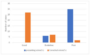
After correction, VA improved in 34 (94.4%) eyes. Of these, 27 (55.5%) eyes had good outcome vision. Six eyes (16.6%) were fully corrected to 6/12. Out of 36 two eyes (5.6%) had poor vision. DISCUSSION Fortunately, visual impairment from uncomplicated aphakia is correctable. The use of spectacles is the oldest means of correcting aphakia and dates back to centuries, but problems of appearance of patients, 35% magnification, ring scotoma, and decrease field of view discouraged its use.6 Contact lenses are another means of correcting aphakia and are superior to spectacles because the retinal image is almost the same size as before cataract surgery. However, because of their thickness, fitting is not easy; they also impede oxygen exchange and are quite expensive. The third means of correction is IOL implantation; this is considered the best method of aphakic visual correction because the implanted lens is almost at its natural anatomic location.6In some countries, aphakia was found to be the major cause of blindness.7 In Our study 3% of the cases are with aphakia after surgery. The lower prevalence might therefore reflect both bias in the sample and a higher income setting than other studies in India and Asia. In a study pooling the results from several Western populations,3the prevalence of unoperated cataract ranged from 15.5% in the 60- to 64-year-old group to 68% in those aged ≥80 and for any aphakia/pseudophakia from 3% to 29% (Table-1). Mean age of patients in present study was 68.9 ± 14.7 years ranging between 35 and 90 years. The mean age showed that aphakia is a condition of the aging population. The reason is most likely due to cataract being a cause of lens removal predominantly among the elderly. We found a19 (52.7%) females and 17(47.2%) males higher prevalence of cataract in women compared with men. This was observed for all types of cataract, both unoperated cataracts, and for all operated cataracts. Women were more likely to have undergone cataract surgery compared with men. Many studies worldwide have reported a higher prevalence of cataract among women. 8,9,10 Nuclear sclerosis is the most common type of cataract, and involves the central or 'nuclear' part of the lens. This eventually becomes hard, or 'sclerotic', due to condensation on the lens nucleus and the deposition of brown pigment within the lens. In its advanced stages. it is called a brunescent cataract. In early stages, an increase in sclerosis cause an increase in refractive index of the lens. Cause of aphakia in present study is with brown cataract(27.8%) followed by pseudoexfoliation(19.4%). Nuclear cataract was higher in north India (48%) compared with south India (38%). For the other types of cataract and for any unoperated cataract, although the differences in the prevalence were significant, the magnitude of the differences was much smaller. The lower prevalence of nuclear cataract in the south might partially be explained by the higher rate of cataract surgery in the south because overall there was no difference between the centres for all unoperated and operated cataract considered together. In present study after correction, VA improved in 34 (94.4%) eyes. Of these, 27 (55.5%) eyes had good outcome vision. Six eyes (16.6%) were fully corrected to 6/12. Out of 36 two eyes (5.6%) had poor vision. About 83% of eyes in this study presented blind, but three eyes of 6 patients presented with borderline vision (6/36 and 6/24). The high rate of blindness is because aphakia causes extremely high hypermetropia. Other studies have reported similar high rate of blindness in uncorrected aphakia.11,12,13 The 6 eyes with borderline presenting vision are most likely myopias because correction of myopia is partly achieved with lens extraction which involves weakening the refractive state of the eye. 31 (86%) eyes were corrected with + 10 D sphere. This finding is consistent or similar with reports from other studies in India and Nepal.14,15 Overall, vision after correction improved in 34 (94%) eyes. In comparison, another study carried out in reported improvement in 66.6% of eyes. Only 27 (55.5%) eyes in this study achieved good (6/18 or better) and 7 (19.4%) borderline vision using +10 D spherical correction.16 This shows that by correcting aphakia with spectacles, contact lens, or IOL implantation, the burden of blindness contributed by aphakia could be reduced by 80.6% in our setting. CONCLUSION Most aphakics are blind, and cataract surgery is the most important causative factor. Corrective measures such as spectacles, contact lenses, and refractive surgery will prevent this blindness. Although the visual outcome was good for most patients in this series, more extended follow-up and a large series of patients are needed to ascertain the effectiveness and safety of this procedure. Aphakia prevention can be achieved by implanting scleral fixation IOLs and AC IOLs and iris claw lenses.
REFERENCES
|
|
 This work is licensed under a Creative Commons Attribution-NonCommercial 4.0 International License.