 Home
Home
Official Journals By StatPerson Publication
|
Table of Content - Volume 12 Issue 2 - November 2019
Incidence and pattern of clinical presentation of ocular injuries due to vehicular accidents
Kavita Patil1*, Rohit Patil2
1Professor and HOD, 2Senior Resident, Department of Ophthalmology, Gulbarga Institute of Medical Sciences, Kalaburgi, INDIA.
Abstract Background: Road traffic accidents (RTAs) involving injury to the eyes and their adnexa incurred in travelling are common. Ocular injuries range from subconjunctival hemorrhage, without much visual significance to sight-threatening injuries. Aim: To document the incidence and pattern of clinical presentation of ocular injuries due to vehicular accidents. Material and Methods: In this prospective study, patients presenting with history of ocular injuries following vehicular accidents were selected. Thorough ophthalmological examination was done and follow up was done after management. Results: Incidence of ocular injuries was 90.4/1000 RTA. Most common ocular manifestation of RTA is subconjunctival haemorrhage 51 (53.65%) followed by ecchymosis 47 (49.44%). The majority of cases 64 (67.39%) had a visual acuity in the range of 6/6-6/12 and only 2 (2.11%) cases had no perception of light after 6 weeks. Conclusion: Ocular injuries due to RTA accounted for 90.4/1000 RTA. Most of the injuries involved the ocular adenexa, which while causing certain degree of cosmetic disfigurement did not lead to any permanent visual sequelae. Key Words: Ocular injuries, road traffic accidents, incidence, subconjunctival haemorrhage, visual acuity
INTRODUCTION An injury to the eye or its surrounding tissues is the most common cause for attendance at an eye hospital emergency department. Road traffic accidents (RTAs) involving injury to the eyes and their adnexa incurred in travelling are common. Ocular injury usually in accidents could be trivial like subconjunctival haemorrhage or simple corneal abrasion to severe such as a contusion leading to fracture of the orbit, concussion or rupture of the globe or perforating injuries due to glass and frequently lead to ocular damage particularly laceration of the eyelids and cheek. Worldwide approximately 1.6 million people are blind from ocular injuries.1 And further 19 million with unilateral visual loss which makes ocular trauma the most common cause of unilateral blindness.1 Severe eye injuries cause a long-lasting disability causing vision loss as well as financial loss to the person and family. Hence, prevention of eye injuries and thereby saving the victim from the disability and stress is the need of the hour. Therefore, the present study was conducted to document the incidence and pattern of clinical presentation of ocular injuries due to vehicular accidents.
MATERIAL AND METHODS In this prospective study, patients presenting with history of ocular injuries following vehicular accidents were selected from the Out-patient Department of the Ophthalmology, Emergency Wards, Casualty of a tertiary care hospital. Inclusion criteria
Exclusion criteria
When a patient presented with a history of ocular injury following vehicular accident, a detailed history regarding age, sex, type of vehicle the patient was travelling and signs and symptoms occurring following the injury were taken.
Depending on the presentation, patients were subjected to detailed examination by ENT surgeon, General surgeon, Maxillo-facial surgeon and General physician. Patients were managed mainly at casualty and OPD level, with some patients admitted for further management and specialized care. Patients were followed up every week after they were discharged from the hospital and the patients who were treated on OPD basis were also followed up every week.
RESULTS Out of 1050 patients of RTA, who presented at our hospital, 95 had ocular injuries. Thus, incidence of ocular injuries was 90.4/1000 RTA. In present study, majority 39 (41.05%) of ocular injuries occurred in the age group of 31-40 years followed by 28.4% in the 41-50 years age group, 20% in 20-30 years age group. The least number of cases 10 (10.52%) were seen over the age of 50 years. The prevalence of ocular injuries was more in males with 67 (70.53%) and 28 (29.5%) in females. The most common 53 (55.76%) of ocular injuries in RTA was due to 2 wheelers. Table 1: Vehicle involved
Right eye was most commonly affected i.e., 52 (54.70%), left eye was involved in 34 (35.8%) cases and both eye involvement was least common i.e., 9 (9.47%).
Table 2: Distribution of cases according to eye affected
Most common ocular manifestation of RTA is subconjunctival haemorrhage 51 (53.65%) followed by ecchymosis 47 (49.44%). Table 3: Pattern of clinical presentation in RTA cases
The majority of cases 64 (67.39%) had a visual acuity in the range of 6/6 – 6/12 and only 2 (2.11%) cases had no perception of light after 6 weeks.
Table 4: Vision at the time of presentation and discharge
Improved visual acuity was seen in 89 (93.72%) cases, in the range between 6/6-6/12 at the end of 6 weeks.
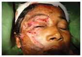
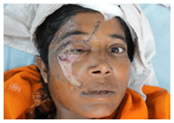
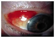
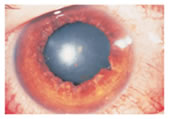
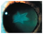
Figure 5 Figure 6 Figure 7 Figure 1: Lid laceration at the presentation; Figure 2: Lid laceration after one week; Figure 3: Subconjunctival haemorrhage Figure 4: Sphincter tear; Figure 5: Berlin’s oedema; Figure 6: Iridodialysis; Figure 7: Traumatic cataract DISCUSSION This study includes a total number of 1050 RTA patients of which 95 patients had ocular injuries. The incidence of ocular injuries in this study was found to be 90.4/1000 RTA. In a study done by Rekhi GS et al,2 ocular trauma accounted for 11.82% of blindness. 119 patients of which 11 were due to road traffic accidents, which accounts for 9.2%. In this study, patients above the age of 18 years were selected. The majority of ocular injuries occurred in the age group of 31-40 years and least number of cases of RTA were seen in patients over 50 years of age. There were a total of 67 males and 28 females. Male:female ratio was 2.4:1. Similar male predominance was found in a study from Western Maharashtra by Patil et al.3 Reddy P also showed a male predominance with a ratio of 2.66:1.4 Out of 95 patients with ocular injuries, most were due to motorized 2 wheelers 53 (55.76%) followed by 4 wheelers 20 (21.04%), pedestrians 15 (15.78%) and 3 wheelers 7 (7.36%). In the study done by Patil et al,3 majority (35%) were due to motorized 2 wheelers followed by 4 wheelers (25.9%). A total number of 11 (11.57%) orbital fractures were seen during the period of our study. Of these 4 cases had medial wall fracture, 2 cases had fracture of orbital rim, 3 cases had floor fractures, one case had lateral wall fracture and 1 patient had a pure blow out fracture. Goswamy et al5 has depicted that out of 12 cases of orbital fracture in 2 years, 5 were due to road traffic accidents. In a study of orbital fractures in a tertiary health care center by Kamath et al,6 22 among 35 patients had infraorbital rim fracture, floor fracture was seen in 10 patients, lateral wall fracture in 4 patients, medial wall fracture in 6 patients, pure blow out fracture in 5 patients and roof fracture in 1 case. Ecchymosis was one of the most common clinical finding; out of the total number of 95 cases, 47 patients had ecchymosis. 46 patients with ecchymosis were associated with other clinical findings while 1 patient had only ecchymosis. In our study, 24 patients had lid tear, most of them being partial thickness lid tear. Four cases had both upper and lower lid tear. 7 cases had associated fracture orbit. Three lid tears had no associated ocular injuries, one patient had Berlin’s oedema and one had vitreous haemorrhage. According to a study by Dabral SM et al,7 who analyzed 82 patients for penetrating eye injuries, concomitant injuries in the eye were laceration of lid (26), lacrimal canalicular injuries (98), conjunctival tears (38), iris tear (30), ciliary body tear (20), lens (95), posterior segment damage (28) and hyphema (32). Subconjunctival haemorrhage was most common clinical finding in our study. 51(53.65%) cases had subconjunctival haemorrhage. They varied from small petechiae to large extravasations. In severe subconjunctival haemorrhage, posterior limit could not be made out. 10 patients did not have any associated clinical findings. Conjunctival tear was seen in 6 (6.31%) patients. Conjunctival tear did not exceed more than 5 mm. Conjunctival foreign body was observed in 8 (8.42%) patients and chemosis in 18 (18.94%) patients. Most of the foreign bodies were sand particles and were lodged in bulbar and upper tarsal conjunctiva. In present study, 16 (16.83%) patients presented with corneal injuries, 3 (3.16%) patients had corneal abrasion, 5 (5.26%) patients had full thickness corneal tear, 3 (3.16%) patients had partial thickness corneal tear and 5 (5.26%) patients had corneal foreign body. 5 patients with full thickness corneal tear were associated with iris prolapse, out of which 1 patient had posterior dislocation of lens and 1 patient had subluxated lens. 2 (2.10%) patients had scleral laceration. None of them were full thickness and were not associated with uveal prolapse. Nanda SK et al8 reviewed 293 patients treated for penetrating ocular injury over a 5 years period and found that 10 (3.4%) were associated with motor vehicle accidents. 6 patients had corneoscleral lacerations, 2 patients each had corneal and sclera lacerations, vitreous surgery was performed in 2 patients, one had intraocular foreign body and one had retinal detachment In our study, 3 (3.16%) cases of hyphaema were presented. All the cases of hyphaema involved <1/3rd of the anterior chamber. All 3 cases had only marginal increase in intraocular pressure. One patient with hyphaema had associated fracture orbit. In a study by Reddy P,4 of 209 cases 59 patients had lid tears, 14 patients had conjunctival tear, 9 patients had corneal tear in which one patient had iris prolapse associated with corneal tear. 6 patients had post segment involvement and 3 patients had hyphema. Amoni SS et al,9 had 76 patients with traumatic hyphema out of which 4 were due to road traffic accidents. Most common findings were traumatic mydriasis 5 (5.26%), 5 (5.26%) cases of Iris prolapse, followed by 3 (3.16%) patients with sphincter tear and 1 (1.05%) with iridodonesis. Our study was compared with a study of 205 cases by Canavan and Archer,10 in which 79 cases had Iris and pupillary injuries. Nine eyes had lenticular involvement. Majority of them 6 (6.31%) had traumatic cataract, 2 (2.10%) had subluxation of lens and 1 (1.05%) had dislocation of lens. 4 (4.21%) patients had early rosette cataract and 2 (2.10%) patients had total cataract. As opposed to 52 eyes out of 212 eyes in a study by Canavan and Archer,10 our study had 3 patients with lenticular damage. Findings were comparable with that of Mukherjee AK et al11 who analyzed 82 patients hospitalized for penetrating eye injuries. Concomitant injuries in the eye were laceration of lid (26), lacrimal canalicular injuries (8), conjunctival tears (38), iris tear (30), ciliary body tear (20), lens (45), posterior segment damage (28), and hyphaema (32). In 12.10% of cases glass caused injury following road traffic accidents. Two patients had vitreous haemorrhage. One patient of vitreous haemorrhage had associated fracture orbit and the other patient had associated ecchymosis and subconjunctival haemorrhage. At the time of presentation both patients had perception of light and improved upto 6/18 and 6/12 respectively after 6 weeks. 2 (2.10%) cases of Berlin’s oedema were observed in this study. Both the cases of Berlin’s oedema had concomitant orbital fracture. Hermman7 found 17 cases of Berlin’s oedema in 677 cases. One (1.05%) patient presented with pre-retinal haemorrhage. The haemorrhage was resolved by 6 weeks with no sequelae. Two patients (2.11%) had traumatic optic neuropathy. Both patients presented with no perception of light, and even after 6 weeks their vision did not improve. Our study was compared with a study of indirect optic nerve injury in two-wheeler riders in North-East India by Bhattacharjee et al12 out of 129 consecutive cases of cranio-orbital injury, 35 patients had indirect traumatic optic neuropathy. Most of the patients with RTA had vision in the range of 6/6-6/12 64 (67.39%) at presentation, and 27 (28.43%) patients had vision in range of 6/18 to counting fingers 1 metre. 2 (2.11%) patients had only perception of light and 2 (2.11%) patients did not have perception of light. At the end of 6 weeks most of the patients i.e. 89 (93.72%) had good vision in the range of 6/6-6/12 and 2 patients, who previously had no perception of light with traumatic optic neuropathy, showed no improvement in visual outcome. In a study by Puzari et al,13 73.33% patient had visual outcome of 6/6, 16.66 % had 6/9-6/60, 6.66 % had CF, 1.66% had PL and 1.66 % had no perception of light. While in study by Shtewi et al,14 30.43 % patient had 6/6, 50.30 % patient had 6/9-6/60, 15.94 % had CF and 3.28 % had no perception of light.
CONCLUSION On conclusion of this study, it was found that ocular injuries due to RTA accounted for 90.4/1000 RTA. Most of the injuries involved the ocular adenexa, which while causing certain degree of cosmetic disfigurement did not lead to any permanent visual sequelae, injuries involving cornea or sclera had bad prognosis and those with optic neuropathy had worst prognosis.
REFERENCES
|
|


 This work is licensed under a Creative Commons Attribution-NonCommercial 4.0 International License.