 Home
Home
|
Table of Content - Volume 13 Issue 1 - January 2020
Role of conjunctival impression cytology in patients with dry eye disease
Rajesh Sharma1, Shaloo Negi2*
1,2Resident, Department of Ophthalmology, Dr. Rajendra Prasad Medical College, Tanda, Himachal Pradesh, INDIA. Email: suhi1551@gmail.com Abstract Background: Conjunctival impression cytology is more sensitive than clinical and laboratory tests, and has the advantage of being able to detect squamous metaplasia before keratinization is clinically detectable. Aim: To study the role of conjunctival impression cytology in dry eye conditions Material and Methods: In this study, total of 142 patients (74 cases and 68 controls) from 20 years up to the age of 60 years, either sex was studied. The Conjunctival impression cytology (CIC) specimens were obtained from the inferior bulbar conjunctiva. It was then stained with Periodic Acid-Schiff and counter stained with Hematoxylin and Eosin. Grading and scoring were carried out using the criteria suggested by Nelson. Results: 45.3% of cases and 15.4% of controls had abnormal cytology. For control group, the mean was 0.84±0.69, for case group, the mean was 1.39±0.787. The difference between cases and controls was statistically significant (p value <0.0001). There was a positive correlation between CIC grades, OSDI scores and BME Grades (correlation coefficient, r >0). Increasing values of OSDI and BME was associated with increasing severity of conjunctival impression cytology. Conclusion: CIC may be predict early changes in ocular surface in dry eye, so that appropriate intervention can be taken before metaplastic changes have taken place. Whether CIC can be used as a first- line investigation for dry eye diagnosis could not be determined. Key words: Dry eye, Conjunctival impression cytology, Nelson grading
INTRODUCTION According to the International Dry Eye Workshop (DEWS 2007), dry eye disease is a multifactorial disease of tears and the ocular surface. Conjunctival impression cytology (CIC) has been widely used as a method for evaluating the ocular surface and superficial cells layers in the diagnosis and follow-up after treatment of several ocular surface tumors of both epithelial and melanocytic origin.1 Impression cytology is a useful non-invasive technique, in which the first or the two outermost layers of the ocular surface epithelium are removed while preserving cellular morphology and studied to determine the state of the conjunctival surface and to classify the severity of squamous metaplasia. This technique allows the investigator to assess epithelial cell morphology, examine cytoplasmic and nuclear characteristics and quantify the goblet cell population in the conjunctiva. Impression cytology is more sensitive than clinical and laboratory tests, and has the advantage of being able to detect squamous metaplasia before keratinization is clinically detectable.2,3 Conjunctival impression cytology has been invaluable as research tool but has not yet become routine diagnostic tool in most clinics because it is relatively cumbersome and time consuming for the clinician and pathologist alike.4 It is minimally invasive, relatively easy to perform and yields reliable information about the area sampled with minimal discomfort to the patient.3,5-7 We intended to study the role of conjunctival impression cytology in dry eye conditions view of overall paucity of data. MATERIAL AND METHODS In this study, total of 142 patients (74 cases and 68 controls) from 20 years up to the age of 60 years, either sex was studied over a period of two years. Informed consent was obtained from all patients prior to enrolment in this study. Inclusion criteria
Exclusion criteria
All the subjects who were included as cases or controls were asked to respond to OSDI questionnaire. Based on their OSDI scores, patients were categorized as having a normal ocular surface (0-12 points) or as having mild (13-22 points), moderate (23-32 points), and severe (33-100 points) ocular surface disease. The participants attaining the score 0-12 were selected as controls while those with score 13-100 were taken as dry eye patients. The enrolled participants were subjected to detailed medical history and clinical examination. Methodology A written informed consent was taken from the participants in their vernacular understood languages (Hindi or English), in the presence of a witness. Demographic information was collected. The patient’s symptoms were thoroughly evaluated and recorded. Detailed slit lamp examination (Slit lamp bio-microscope- Haag Streit BM 900) was done to exclude any lid conditions such as meibomitis, any eye lid deformities and lagophthalmos which may disturb the normal tear film. The Conjunctival impression cytology (CIC) specimens were obtained from the inferior bulbar conjunctiva using a circular 0.22-micron pore size Millipore mixed cellu¬lose acetate filter paper patch, 13 mm in diameter cut into two halves. The eye was anesthetized using one drop of 4% Xylocaine. The lacrimal lake at the inner canthus was dried with a cotton tip applicator. The filter paper was grasped with a blunt smooth edge forceps and applied to the inferior bulbar conjunctiva. The filter paper was pressed gently with an applanation head piece of Goldman’s applanation tonometer held in the other hand. After 4 to 10 seconds, the paper strip was removed in a peeling fashion and the specimen was transferred to the glass slide coated with albumin paste by pressing the filter paper on it. The slide was labelled and numbered. The slide was put into Koplin jar containing fixative solution made up of Glacial acetic acid, Formaldehyde and Ethyl Alcohol in a 1:1:20 volume ratio. The slide was kept in fixative solution for 10 minutes and transported to the Department of Pathololgy. It was then stained with Periodic Acid-Schiff and counter stained with Hematoxylin and Eosin. Grading and scoring were carried out using the criteria suggested by Nelson. Grades 0 and 1 were regarded as normal, whereas grades 2 and 3 were considered to represent abnormal cytology.
Figure 1: Conjunctival impression sample being taken
The Biomicroscopic Examination (BME) of the meibomian glands, lids, conjunctiva and tear film was performed at a slit lamp to grade the presence/severity of meibomian gland disease (MGD) and to assess signs of ocular surface abnormality and inflammation. The Tear film Break-Up Time (TBUT) assessment was done and the readings analysed. The Schirmer’s I test was done and the reading noted. Statistical analysis The data was coded and compiled on Microsoft Excel spread sheet. Categorical data was expressed in terms of rates, ratios and percentages. Continuous variables were expressed as mean ± standard deviation(SD). The data was analysed by test of proportion and chi-square test. A probability value (‘p’ value) of <0.05 was considered as statistically significant.
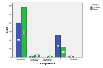
RESULTS A total of 142 subjects (74 cases and 68 controls) from 20 years of age to 60 years were included in the study. There were 51 males (35.9%) and 91 females (64.1%), with male to female ratio 1:2. There was a preponderance of females in our study in both the groups. However, the difference between males and females in different groups was not statistically significant (p=0.840). Mean age of the participants was 38.61±12.67 (minimum 20 years and maximum 60 years of age) in cases and 36.12±12.21 (minimum 20 years and maximum 60 years of age) in controls. In the present study, 32.4% cases of dry eye were in age range of 20-30 years, 27.5% in 31-40 years, 22.5% in 41-50 years and 17.6% in 51-60 years. Conjuntival congestion was seen in 78.4% of cases and 58.8% of controls. Similarly, Normal conjunctiva was seen in 16.2% of cases and 38.2% of controls. The difference was significant (p< 0.05).
Graph 1: Conjunctival Finding in Cases and Controls
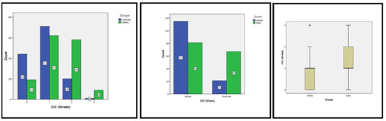
For CIC test, by taking grade 0-1 as normal and grade 2-3 as abnormal cytology, 54.7% of cases and 84.6% of controls had normal cytology. Similarly, 45.3% of cases and 15.4% of controls had abnormal cytology. For control group, the mean was 0.84±0.69, for case group, the mean was 1.39±0.787. The difference between cases and controls was statistically significant (p value <0.0001).
Graph 2 Graph 3 Graph 4 Graph 2: Cases and Controls with Respective CIC Grades; Graph 3: Normal and Abnormal CIC Grades for Cases and Controls; Graph 4: Mean and Standard Deviation of CIC Grades for Cases and Controls
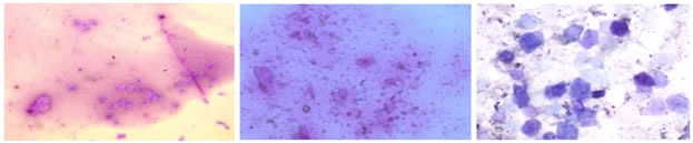
Fig 1 (a, b, c): Histological slides showing Nelson’s classification grades 1-3 a. Nelson’s classification –Grade 1 - Round to polygonal cells with relatively raised N/C ratio and moderate number of goblet cells b. Nelson’s classification- Grade 2 - Polygonal squamous epithelial cells with relatively raised N/C ratio and lesser number of goblets cells c. Nelson’s classification – Grade 3- Polygonal cells with pyknotic nucleus. No goblet cells It was observed that 61.4% of subjects with abnormal impression cytology (grade 3,4) had Schirmer values >15mm, while 79.6% of subjects with normal impression cytology(grade 0,1) had Schirmer values >15mm. Similarly, it was found that 17.1% of subjects with abnormal cytology had Schirmer scores <5mm in comparison to 3.6% of subjects with normal cytology. The difference in both the groups was found to be statistically significant(p <0.0001). It was observed that 25% of subjects with abnormal impression cytology(grade 3,4) had TBUT values >10 sec, while 65.8% of subjects with normal impression cytology(grade 0,1) had TBUT values >10 sec. Similarly, it was also found that 37.5% of subjects with abnormal cytology had TBUT scores <5 sec in comparison to 6.5% of subjects with normal cytology. The difference in both the groups was found to be statistically significant (p <0.0001). It was observed that 52.3% of subjects with abnormal impression cytology (grade 3,4) had normal BME grade score (grade 0,1), while 77.6% of subjects with normal impression cytology (grade 0,1) had normal BME grade score (grade 0,1). Similarly, it was also found that 47.7% of subjects with abnormal impression cytology had abnormal BME grade score (grade 2,3) in comparison to 22.4% of subjects with normal impression cytology. The difference in both the groups was found to be statistically significant (p <0.0001). There was a positive correlation between CIC grades, OSDI scores and BME Grades (correlation coefficient, r >0). Increasing values of OSDI and BME was associated with increasing severity of conjunctival impression cytology. There was a negative correlation between CIC grades, Schirmer values and TBUT scores (r <0). The decreasing level of TBUT and Schirmer were associated with increasing severity of conjunctival impression cytology
DISCUSSION Goblet cells are known to secrete soluble mucins into the tear film, playing a major role in the defence and regulation of the ocular surface. Since lack of goblet cells is one of the hallmarks of all dry eye syndromes in particular mucin deficiency. The classical impression cytology approach which allows the calculation of goblet cell density and the staging of squamous metaplasia was not used in our study, as goblet cell density was not determined due to procedural limitations. Instead, the Nelson grading system was used as used by Shrestha E et al8 in their study. In our study, out of 74 cases, 45.34% had abnormal impression cytology; amongst these (39.2 % had Nelson grade 2 and 6.1% Nelson grade 3 changes, respectively); whereas amongst 68 controls, only 15.4% (Nelson grade 2 and 3) had abnormal cytology and 84.6% had normal cytology (Nelson grade 0 and 1). These results were similar to those obtained by Shrestha E et al8 where of 114 dry eye cases, 49.2% eyes showed decreased or absent goblet cells. In 72 normal individuals 73.7% eyes showed normal goblet cells and 26.3% of eyes showed decreased or absent goblet cells (p < 0.001). The difference observed in our study in controls with normal cytology in comparison to those in the above mentioned study could be attributed to the criteria used in selection of controls, which was OSDI in our study and TBUT, Schirmer and symptomatology in their study. In a study by Kumar P et al,9 they found that 46% cases of dry eye had abnormal cytological changes (Nelson grade II-III), whereas this type of cytology was observed in only 32.8% of controls. Similar results were observed in our study except that controls with abnormal cytology were 15.4% in our study not 32.8% as in their study. This could be due to selection criteria used in our study was OSDI score <12 for controls. The OSDI questionnaire was used because it reliably assesses the severity, natural history, and effects of dry eye. Compared to other questionnaires, the OSDI has a sensitivity of 60% and specificity of 79%105. In their study, Kumar P et al9 used only eight symptoms for diagnosing dry eye symptomatically. In a study by Bhargava et al113 it was observed that Impression cytology was normal (Nelson Grades 0 and 1) in 46.4% cases. Nelson grade 2 changes predominated (43%) amongst those having abnormal CIC (53.6%), which was comparable to our study in which 54.7% of cases had normal cytology and Nelson grade 2 changes predominated (39.2%) amongst those having abnormal CIC (45.3%). On the contrary, 6.1% controls had abnormal cytology in their study which was 15.4% in our study. Bhargava et al10 observed that symptom based assessment needed to be combined with tests like CIC to increase diagnostic accuracy and to effectively monitor response to treatment. They concluded that dry eye scoring system, CIC, and TBUT correlate well and may hold good diagnostic accuracy, may detect early dry eye changes, when used in combination for diagnosis of dry eye in computer users. In another study by Bhargawa et al,11 the diagnostic accuracy of CIC, TBUT, Schirmer and RBS in dry eye patients was evaluated and compared with age and sex matched controls. The area under the curve(AUC) was measured using receiver-operating characteristics (ROC) curve. The diagnostic accuracy was CIC (AUC=0.957) >TBUT (AUC=0.793) >Schirmer (AUC=0.765) >RBS (AUC=0.723). One of the most important feature of dry syndrome is alteration of conjunctival and corneal epithelium as seen on vital staining (Rose Bengal and Lissamine green). However, these methods are less sensitive and specific, have low diagnostic accuracy, do not indicate degree of squamous metaplasia or changes in goblet cell density and do not correlate with disease severity.12 Correlation analysis (with regression) suggests that there was a significant correlation of dry eye symptoms with Nelson grade. Thus, the main advantage of CIC may be predicting early changes in ocular surface in dry eye (undetected by routine tear function tests), so that appropriate intervention can be taken before metaplastic changes have taken place.
CONCLUSION CIC may be predict early changes in ocular surface in dry eye (undetected by routine tear function tests), so that appropriate intervention can be taken before metaplastic changes have taken place. Although CIC correlates best with dry eye symptoms, the equipment needed to carry out the testing may not be readily available in all settings. Whether CIC can be used as a first-line investigation for dry eye diagnosis could not be determined.
REFERENCES
Policy for Articles with Open Access
|
|

 This work is licensed under a Creative Commons Attribution-NonCommercial 4.0 International License.