 Home
Home
|
Table of Content - Volume 13 Issue 1 - January 2020
A hospital based study of tear film break up time in patients with dry eye disease
Rajesh Sharma1*, Shaloo Negi2
1,2 Resident, Dr. Rajendra Prasad Medical College, Tanda Himachal Pradesh, INDIA. Email: suhi1551@gmail.com
Abstract Background: Dry eye disease is a multifactorial disease of tears and the ocular surface. A combination of various subjective and objective measurements is often used to determine the presence and/or severity of dry eye in an individual. Aim: To study the tear film Break up time in patients with dry eye disease. Material And Methods: A total of 142 patients (74 cases and 68 controls) were studied over a period of two years. The Tear film Break-Up Time(TBUT) assessment was done and the readings analysed. The Schirmer’s I test was done and the reading noted. Results: It was observed that 55.4 % eyes of cases and 37.5% eyes of controls had TBUT value of =<10 sec, similarly 44.6% of cases and 62.5% of controls had values>10 sec. The sensitivity, specificity, PPV and NPV of TBUT test were 75%, 65.8%, 49.6% and 85% respectively. Conclusion: The study of tear film break up time is an important step in the diagnosis, management and prognosis of dry eye. The interpretations of this test are easy and therefore, this test should be undertaken as a routine in the early diagnosis of dry eye syndrome. Key Words: Dry eye, tear film break up time, Schirmer’s I test
INTRODUCTION The ocular surface functions as a unit and as such can be deleteriously affected by a wide range of pathologies, adversely affecting any of its underlying structures. This can lead to tear film abnormalities, inflammatory changes, neural abnormalities or simply produce symptoms indistinguishable from dry eye disease. Dry eye disease is a multifactorial disease of tears and the ocular surface. It results in symptoms of discomfort, visual disturbances and tear-film instability with potential dam¬age to the ocular surface. It is accompanied by increased osmolarity of the tear film and inflammation of the ocular surface.1 A combination of various subjective and objective measurements is often used to determine the presence and/or severity of dry eye in an individual.2,3 In addition, it has been recognised, particularly in moderate/mild dry eye, that diagnostic tests are prone to disagree and give conflicting results. Begley et al4 previously reported that ocular surface staining did not always correlate with patient’s dry eye symptoms, while Kallarackal et al5 found a poor correlation between Schirmer’s test and tear break up time in dry eye patients. This hospital based study was conducted to study the tear film Break up time in patients with dry eye disease.
MATERIAL AND METHODS A total of 142 patients (74 cases and 68 controls) were studied over a period of two years. Informed consent was obtained from all patients prior to enrolment in this study. Inclusion criteria
Exclusion criteria
All the subjects who were included as cases or controls were asked to respond to OSDI questionnaire. Based on their OSDI scores, patients were categorized as having a normal ocular surface (0-12 points) or as having mild (13-22 points), moderate (23-32 points), and severe (33-100 points) ocular surface disease. The participants attaining the score 0-12 were selected as controls while those with score 13-100 were taken as dry eye patients. The enrolled participants were subjected to detailed medical history and clinical examination. Methodology Detailed slit lamp examination (Slit lamp bio-microscope- Haag Streit BM 900) was done to exclude any lid conditions such as meibomitis, any eye lid deformities and lagophthalmos which may disturb the normal tear film. The Tear film Break-Up Time(TBUT) assessment was done and the readings analysed. The tear film break-up time was defined as the interval between the last complete blink and the first appearance of a dry spot, or disruption in the tear film. A sterile fluorescein strip wetted with a drop of normal saline was instilled onto the bulbar conjunctiva as follows: with fixation directed inferonasaly, and the upper lid gently retracted the fluorescein strip was introduced at an approximate 30° angle to the superior temporal bulbar conjunctiva and touched for 1-2 seconds, so that 1-2 mm of the flat side makes contact. The patient was instructed to blink naturally, without squeezing, several times to distribute the fluorescein. Within 10 - 30 seconds of the fluorescein instillation, the patient was asked to stare straight ahead without blinking, until told otherwise. Slit-lamp magnification was set at 10X. Cobalt blue light was used for observing the tear film over the cornea. Stopwatch was used to record time between last complete blink and first appearance of dry spot. Once TBUT was observed, patient was instructed to blink freely. Two readings were taken and averaged. The Schirmer’s I test was done and the reading noted. The test was done by placing the Schirmer’s strip, made up of Whatman filter paper number 41 with dimensions 5mm X 35mm. The initial 5mm of the strip was folded and kept in junction of lateral one third and medial two third of the lower fornix of the eye. It was kept for 5 minutes. The wetting of the strip at the end of 5 minutes was noted using the scale present on the strip. Statistical analysis The data was coded and compiled on Microsoft Excel spread sheet. Categorical data was expressed in terms of rates, ratios and percentages. Continuous variables were expressed as mean ± standard deviation(SD). The data was analysed by test of proportion and chi-square test. A probability value (‘p’ value) of <0.05 was considered as statistically significant.
RESULTS Out of 142 subjects (74 cases and 68 controls), 51 were males (35.9%) and 91 were females (64.1%), with male to female ratio 1:2. There was a preponderance of females in our study in both the groups. However, the difference between males and females in different groups was not statistically significant (p=0.840). Mean age of the participants was 38.61±12.67 (minimum 20 years and maximum 60 years of age) in cases and 36.12±12.21 (minimum 20 years and maximum 60 years of age) in controls. In the present study, 32.4% cases of dry eye were in age range of 20-30 years, 27.5% in 31-40 years, 22.5% in 41-50 years and 17.6% in 51-60 years.
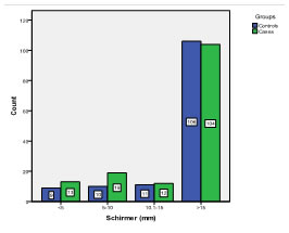
Graph 1: Schirmer’s Test Values for Cases and Controls Schirmer’s I test was done without anaesthesia in cases and controls. For control group, the mean was 24.32±10.088 in mm, 95% Confidence Interval for Mean lower bound was 22.61 and upper bound 26.03. For case group, the mean was 23.34±11.662 in mm, 95% Confidence Interval for Mean lower bound was 21.44 and upper bound 25.23. The difference was found to be not significant (p=0.379).
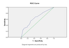
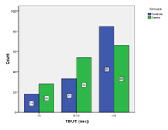
Graph 2: ROC Curve for Schirmer’s Test For TBUT test, value of 10 sec was taken as cut-off for diagnosing a case of DED. It was observed that 55.4 % eyes of cases and 37.5% eyes of controls had TBUT value of =<10 sec, similarly 44.6% of cases and 62.5% of controls had values>10 sec. Graph 2: Cases and Controls with respective cut off TBUT values
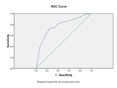
For control group, the mean was 12.33± 5.795 in seconds and for case group, the mean was 10.05±5.498 in seconds. The difference was found to be statistically significant (p value <0.05). In our study, sensitivity, specificity, PPV and NPV of TBUT test were 75%, 65.8%, 49.6% and 85% respectively. Graph 4: ROC Curve for TBUT
DISCUSSION In our study, Schirmer’s I test was done without anaesthesia in cases and controls. For control group, the mean was 24.32±10.088 in mm, for case group, the mean was 23.34±11.662 in mm. The difference was found to be not significant(p=0.379). Although performing the Schirmer’s I test with anaesthetic may provide a more accurate picture of basal secretion, the utility and overall effectiveness of anaesthetic administration in conjunction with the Schirmer is controversial.6 Use of anaesthetic has been implicated in disruption of cell junctions which may increase surface staining leading to erroneous conclusions during the evaluation of surface integrity.7 As corneal and/or conjunctival staining is routinely performed after the Schirmer’s test, the inclusion of anaesthetic may inadvertently result in misclassification of the presence and/or severity of dry eye.8 Many reviews and research papers have documented high variability, low reproducibility and poor correlation with other signs and symptoms of dry eye.5,9,10,11 In an attempt to minimize variability, numerous variations of the Schirmer’s test (in addition to anaesthetic use), were proposed including closing the eyes, using dim light, reducing the test time and the use of different filter materials. But no such alteration in methodology has resulted in a consistent improvement in Schirmer reproducibility or diagnostic sensitivity. Furthermore, the range of values is such that regardless of a cut-off point, false negative and/or positive identification of subjects as DED patients is common.6 Danjo et al12 did a study to evaluate diagnostic usefulness and cut- off value of Schirmer's I test in the Japanese diagnostic criteria of dry eye. 100 eyes of 50 patients with Sjogren's syndrome underwent a series of diagnostic tests, including Schirmer's I test, cotton thread test, TBUT, fluorescein staining, and Rose Bengal staining. They were classified into definite dry eye, probable dry eye, and normal eye according to the Japanese criteria. The diagnostic usefulness of Schirmer's I test was evaluated in comparison with that of the cotton thread test or TBUT, based on the diagnostic outcome by combination of the individual tests plus vital staining tests. The cut-off value of Schirmer's I test was evaluated, based on the results of sensitivity and specificity rates at each cut-off value from 0 to 10 mm. The diagnostic usefulness of Schirmer's I test was inferior to that of TBUT, but superior to that of cotton thread test. The sensitivity and specificity were 88% and 35% respectively, at a cut-off value of 10 mm. In another study by Kumar et al,13 the sensitivity and specificity were found to be 65.69% and 68.5% respectively. Rahul Bhargava and Prachi Kumar10 found the sensitivity and specificity of Schirmer’s test as 38.2% and 82% respectively. The sensitivity and specificity observed in our study, taking cut-off value as 10mm, were 30.7% and 87.8% respectively, which was comparable to that was found by Rahul Bhargava and Prachi Kumar. Hence, it can be concluded that due to high variability, low reproducibility and poor correlation with other signs and symptoms of dry eye, Schirmer’s test is unreliable and false negative and/or positive identification of dry-eyed subjects is common. For TBUT test, value of 10 sec was taken as cut-off for diagnosing a case of DED. It was observed that 55.4 % eyes of cases and 37.5% eyes of controls had TBUT value of =<10 sec, similarly 44.6% of cases and 62.5% of controls had values>10 sec. For control group, the mean was 12.33±5.795 in seconds and for case group, the mean was 10.05±5.498 in seconds. The difference was found to be statistically significant (p value <0.05). In our study, sensitivity, specificity, PPV and NPV of TBUT test were 75%, 65.8%, 49.6% and 85% respectively. Our results were similar to that of study by Kumar et al13 where sensitivity, specificity and PPV were observed as 80.43%, 64.96% and 45.4% respectively. In another study by Rahul Bhargava and Prachi Kumar,10 sensitivity and specificity were noted as 88.6% and 82.4% respectively. In a study by Danjo et al12 the diagnostic usefulness of TBUT was found to be superior to that of Schirmer's I test, which was similar to our study. In a study by Baudouin et al,14 TBUT was considered to have a specialized role in the diagnosis and evaluation of DED. TBUT was a routine test for tear instability, and the panel agreed that it was essential for confirming/verifying diagnosis of dry eye in cases of a high symptomatology score. The assessment of tear film stability using fluorescein is considered by many to be the most important clinical diagnostic test available.15,16 with previous studies demonstrating tear break up time to be reduced in nearly all forms of dry eye, from KCS to meibomian gland disorders.17-19 However, as a clinical tool, TBUT using fluorescein has been criticised due to concerns about its reproducibility and variability between normal subjects on different days.20
CONCLUSION It can be safely concluded that the study of tear film break up time is an important step in the diagnosis, management and prognosis of dry eye. The interpretations of this test are easy and therefore, this test should be undertaken as a routine in the early diagnosis of dry eye syndrome.
REFERENCES
Policy for Articles with Open Access |
|
 This work is licensed under a Creative Commons Attribution-NonCommercial 4.0 International License.