 Home
Home
|
Table of Content - Volume 13 Issue 3 - March 2020
A study on success rates in no glue no suture technique of pterygium excision with conjunctival autograft in rural population of Chittoor district, Andhra pradesh, India
Divya D1, N Sathish Kumar2, Hanumanthappa3, Vishnu Priya4, Jayasimha5
1Assistant Professor, 2Associate Professor, 3Professor & HOD, 4Senior Resident, 5Junior Resident, Department Of Ophthalmology, Apollo Institute of Medical Sciences and Research, Murakambattu, Chittoor District, Andhra Pradesh INDIA. Email: eyesurgeon2004@gmail.com
Abstract Background: Pterygium is a fibroelastotic degeneration of the subconjunctival tissues which proliferate as vascularized granulation tissue to invade the cornea. Symptomatic pterygium needs to be excised. But pterygium surgery remained an enigma through ages. As Bare scleral excision has a disadvantage of recurrence, conjunctival autografting is done nowadays. Materials and Methods: The present study is a retrospective study conducted at the teaching hospital, Chittoor district during the period of November 2018 to November 2019. The study included 107 patients with Progressive Pterygium, who underwent surgery for the same. Data were collected from the hospital records. Results: Out of 107 patients, who underwent surgery, 76 (71%) were female and 31 (29%) were male. Most of the patients were aged between 41-50 years (39.3%). Nasal pterygium was seen in 100 (93.5%), Temporal pterygium was seen in 7 (6.5%) patients. Of 107 patients only 5 (4.6%) patients had bilateral pterygium whereas unilateral pterygium was seen in 102 (95.4%) patients. During postoperative follow-up, post-op complications were seen in only 10 (9.4%) patients, which included Graft displacement in 5 (4.7%) patients during the first week following surgery, Granuloma formation in 2 (1.9%) patients after 2 weeks of surgery, recurrence of pterygium in 3 (2.8%) patients during 2 months postoperatively. Graft rejection was not observed in any of the patients. Conclusion: Conjunctival autografting with Autologous serum was found to be a more effective method following pterygium excision. The main advantage we observed is the chance of Graft rejection and transmission of diseases like HIV and Hepatitis B were not there. This Technique got an easy learning curve for any Ophthalmic surgeon. It demands no extra cost but provides all the benefits of fibrin glue without its drawbacks and without suture related postoperative complications. It is very economical to use Autologous serum for conjunctival autografting following Pterygium excision for all the patients irrespective of their affordability. Key Word: pterygium excision.
INTRODUCTION Pterygium, is a wing-shaped, fibrovascular overgrowth arising from subconjunctival tissue extending across the limbus onto the cornea, destroying superficial layers of stroma and Bowman's membrane, the whole being covered by conjunctival epithelium 1,2. The prevalence rate of primary pterygium varies from 0.7% to 31% in various populations around the world 3 Although exact etiology is not known, working outdoors increase the risk 1.5-fold and the other risk factors include genetic predisposition, chronic environmental irritations such as dust, dryness, heat, and ultraviolet rays.4,5 The excision of pterygium with no added therapy (Bare sclera technique) was widely practiced because it was believed to be safe and simple. However, with time it became apparent that the recurrence rate was unacceptably high ranging from 32% to 88% 6. Several methods were implemented with the aim of improving the success rates, among them conjunctival autograft was one of the recently recommended techniques. Conjunctival autografting can be done by using either Sutures or Fibrin glue or Autologous serum. This study concerns itself to the success rates in pterygium excision with Conjunctival autografting without using any sutures and Fibrin glue but using patients' autologous serum for attaching the graft to bare sclera following pterygium excision.
MATERIALS AND METHODS The present study is a retrospective study conducted at a Teaching hospital, Chittoor district during the period of November 2018 to November 2019. The study included 107 patients with progressive Pterygium, who were operated for the same. The data was collected from hospital records. The diagnosis of Progressive pterygium was done clinically. A thorough clinical assessment of these patients was done by taking a brief history of presenting complaints, family history, and then thorough examination was done, which included Assessment of visual acuity through Snellen’s chart, slit-lamp examination. Fundus evaluation, as a routine was done by direct and indirect ophthalmoscopy. At the time of surgery, Consent was taken from the patients for surgery as well as special consent was taken from the patient for using the data for future publications. Inclusion criteria:
Exclusion criteria:
A written informed consent explaining the complications and possibility of recurrence was obtained from all patients. And special consent was taken regarding their acceptance for publication in any journal in the future. Preoperative preparation: Patients were started on,
Surgical Procedure followed was:
RESULTS Out of 107 patients, who underwent surgery, 76 (71%) were female and 31 (29%) were male. Most of the patients were aged between 41-50 years (39.3%). Nasal pterygium was seen in 100 (93.5%) patients, whereas temporal pterygium was seen in only 7 (6.5%) patients. Of 107 patients, only 5 (4.6%) patients had bilateral pterygium whereas unilateral pterygium was seen in 102 (95.4%) patients. During postoperative follow-up, post-op complications were seen in only 10 (9.4%) patients of which Graft displacement was noted in 5 (4.7%) patients during the first week following surgery, Granuloma formation was seen in 2 (1.9%) patients after 2 weeks of surgery, recurrence of pterygium was seen in 3 (2.8%) patients during 2 months postoperatively. Graft rejection was not observed in the patients during 6 months of postoperative follow up TABLE 1: GENDER DISTRIBUTION
TABLE 2: AGE OF PRESENTATION
TABLE 3: LOCATION OF PTERYGIUM
TABLE 4: LATERALITY OF PTERYGIUM
TABLE 5: POSTOPERATIVE COMPLICATIONS
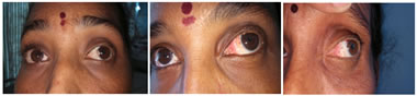
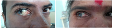
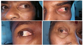
Figure 1: Conjunctival autografting with Autologous serum (Nasal Pterygium before surgery, 1 week Post-op, 1 month Post-op) Figure 2: Conjunctival autografting with Autologous serum Figure 3: Conjunctival autografting with Autologous serum DISCUSSION Pterygium is a fibroelastotic degeneration of the subconjunctival tissue which proliferates as vascularized granulation tissue to invade the cornea. If not treated it may encroach the entire pupillary axis and thus cause a significant decrease in the visual acuity. The contractile forces of pterygium on the peripheral horizontal cornea leads to significant flattening of the horizontal meridian (with the rule astigmatism), which is proportional to the size of the pterygium7,8,9 Other symptoms being redness, foreign body sensation, diplopia, cosmetic disfigurement. There are several surgeries for pterygium of which Bare scleral excision is the basic and simple method of pterygium excision. But it is no longer acceptable in the treatment of pterygium as the chances of recurrence are more common10,11. The reported rates of recurrence are 39% 12. So, Pterygium surgery remained an enigma through ages. Several studies were conducted to modify the bare sclera excision to conjunctival autografting. This has an acceptable low recurrence rate, good cosmesis, and no gross post-operative complication11,13,14,15 Conjunctival autografting can done following Pterygium excision in 3 different ways. One way is by suturing the graft to the bare sclera by using either 10-0 Nylon or 7-0 vicryl suture material. Another way of securing conjunctival autograft is by using Fibrin glue. Another way of conjunctival autografting is by using Autologous serum. Sutures may cause patient discomfort, chronic inflammation, granuloma formation symblepharon formation 16,17, ocular motility restriction, diplopia, scleral necrosis, and infection18,19. The presence of sutures may lead to prolonged wound healing and fibrosis 20. Grafting by using Fibrin glue is easier with less surgical time and short postoperative recovery time and fewer chances of postoperative complications and recurrence. But Fibrin glues are currently manufactured from human plasma and therefore carry the theoretical risk of transmissible diseases like prion disease, HIV, Hepatitis A virus and parvovirus B19 14,21. It carries the potential risks of anaphylaxis. Another drawback with Fibrin glue is due to its higher cost and difficulty in availability. Fibrin glue is bio-degradable within 3 hours of preparation, which demands a certain amount of quick positioning and alignment of the graft. Securing the conjunctival autograft to bare sclera using autologous blood is a new approach, also known as “suture and glue-free autologous graft.” This procedure has excellent results without any complications associated with sutures and fibrin glue. Recurrence rate following this technique was around 2.6% 22, with the mean surgical time of 11 min, no graft rejections were noticed, and none of the pterygium recurred 23 Following conjunctival autografting with autologous serum, the apposition of the lids to the bulbar conjunctiva provides a natural biological dressing and confers a unique wound-healing environment. Apart from a physical barrier, the lids provide compression, a smooth frictionless surface, and avascular bed with immune capability in close proximity to the injury site
CONCULSION The main advantage we observed through this technique is the absence of Graft rejection, less surgical time, low recurrence rate and less postoperative complications. Unlike Fibrin glue, chances of transmission of diseases like HIV and Hepatitis B are less .This Technique got an easy learning curve for any Ophthalmic surgeon. It demands no extra cost but provides all the benefits of fibrin glue without its drawbacks and without suture related postoperative complications. It is very economical to use Autologous serum for conjunctival autografting following pterygium excision for all the patients irrespective of their affordability.
REFERENCES Policy for Articles with Open Access
|
|
 This work is licensed under a Creative Commons Attribution-NonCommercial 4.0 International License.