|
Table of Content - Volume 14 Issue 2 - May 2020
A study of comparison of intraocular pressure in diabetics and non-diabetics in a rural area
Pallavi Bangalore Acharlu1*, Sujatha Vijayalekshmi2, Vijay Kumar Srivastava3
1Assistant Professor, 2Professor, 3Professor and HOD, Department of Ophthalmology, MVJ Medical College & Research Hospital, Hoskote, Bangalore, Karnataka, INDIA. Email:pallaviacharlu@gmail.com
Abstract Background: Diabetic Retinopathy and Glaucoma are the leading causes for irreversible blindness worldwide. Few studies show positive correlation with Intra Ocular Pressure (IOP) and Diabetes. Therefore our study was conducted to check the association of Intraocular Pressure in patients with Diabetes and patients without Diabetes. Methods: Our study is a hospital based cross-sectional study. A complete examination of anterior segment was done using slit lamp. Posterior segment was examined using Direct and Indirect Ophthalmolscope. Intraocular pressure was recorded using a Perkins Tonometer. Results: 120 patients were included in our study who fall into the inclusion criteria. The mean Intraocular pressure in Diabetic patients was 13.53 ± 1.90 mm of Hg and mean IOP in Non Diabetic patients was 11.92 ± 1.17 mm of Hg. Diabetic patients had significantly higher IOP than the Non Diabetic patients (p<0.0001).Conclusion: Our study showed significantly higher intraocular pressures in patients with Diabetes than without Diabetes. This would suggest that Diabetics should be monitored regularly for intraocular pressure to detect an early onset of Glaucoma in susceptible patients. Key Words: Diabetes Mellitus, Diabetic Retinopathy, Intraocular pressure, Glaucoma.
INTRODUCTION Intraocular fluids exert some pressure on the walls of the eyeball to maintain the rigidity of the eye. This pressure exerted is called as Intraocular pressure (IOP).The normal values of intraocular pressure is 10-21mm of Hg. The dynamic equilibrium between the production and drainage of the aqueous humour is required to maintain the normal level of IOP. Various local factors which influence the IOP are rate of formation of aqueous humour, resistance to outflow of aqueous humour, Episcleral venous pressure, pupil size and refractive status of the eye. Various general factors like age, sex, heredity, blood pressure, body mass index, exercise etc may influence the level of intraocular pressure.1Intraocular Pressure is one of the most important risk factor for Glaucoma, and is often seen in association with Diabetes Mellitus and Systemic Hypertension2. Due to day by day increase in the prevalence of Diabetes Mellitus, it has become a major health problem in India. The various organs which will be damaged in association with Diabetes Mellitus are Eye, Kidney, Heart, Blood vessels and Nerves. Diabetes Mellitus affects eyes and had various complications, therefore it acts as important risk factor for Diseases of eye.3,4 The various ocular complications caused by Diabetes are Diabetic Retinopathy, Cataract (opacification of the lens), Cranial nerve palsies, Rubeosis iridis and Glaucoma (raise in intraocular pressure causing damage to the optic nerve)4.Though the mechanism for raise in intraocular pressure is not clear, Genetic, autonomic dysfunction and Osmotic diffusion are various the postulated etiologies5. Various studies have found correlation between Diabetes and Glaucoma 6-10, however few studies have failed 11-17. Therefore with the intention of finding the association between IOP and Diabetes, we conducted this study. We examined the IOP in individuals with diabetics and correlated with Non Diabetic individuals.
MATERIALS AND METHODS Aim: To determine the correlation between Intraocular pressure in Diabetic patients and Non Diabetic patients. Study design: It is a hospital based cross-sectional study conducted with patients presenting to the Ophthalmology Outpatient Department (OPD) from September 2019 to Dec 2019 at MVJ Medical College and Research Hospital, Hoskote. Informed consent was taken from the patients for this study. 120 patients (240 eyes) were included in this study. Inclusion criteria:
Exclusion criteria:
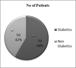
The demographic data of the patients like, name, age, gender, address and occupation was taken. Later detailed medical, past, systemic, surgical, treatment history were recorded. After this ocular examination was done which included visual acuity, Retinoscopy and Refraction, detailed slit lamp evaluation of anterior segment, examination of fundus using Direct and Indirect Ophthalmoscope. Intraocular pressure readings were taken using Perkins Tonometer. Among the Diabetic patients the patients were categorized into 5 groups according to the fundus finding as Group1 -No Diabetic Retinopathy, Group2-Mild Non Proliferative Diabetic Retinopathy (Mild NPDR), Group3- Moderate Non Proliferative Diabetic Retinopathy (Moderate NPDR), Group4-Severe Non Proliferative Diabetic Retinopathy (Severe NPDR) and Group5- Proliferative Diabetic Retinopathy (PDR).Gonioscopy and Perimetry was planned if IOP>21mm Hg or optic disc changes suggestive of Glaucoma was present. Statistical Data Analysis:All the findings were entered in excel sheet. They we expressed as Mean with ± standard deviation. Comparison of Mean Intraocular pressure of Diabetic patients group and Non Diabetic patients group was done by using unpaired student t test. For the comparison between different groups for continuous variables Analysis of Variance (ANOVA) was used.RESULTS 120 patients were selected for the study. Out of 120 patients 70 (140 eyes) were Diabetics and 50 (100 eyes) were Non Diabetics (Fig1). The mean (±standard deviation) age of Diabetic patients group was 55.06 ± 9.70 years (Range 40 to 75 years). Out of 70 Diabetic patients 54 were Males (age 55.26 ± 9.62 years) and 16 were Females (age 54.38 ± 10.26 years). The mean age of the Non Diabetic patients was 53.52 ± 8.81 years .Out of 50 Non Diabetic patients 35 were Males (age 53.49 ± 8.81 years) and 15 were Females (age 53.60 ± 9.11 years) Table 1.
Figure 1: Diabetics and Non-Diabetics among study population Table 1: Distribution of Age and gender in the study Groups
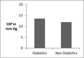
The mean Intraocular pressure in Diabetic patients was 13.53 ± 1.90 mm of Hg and Non Diabetic patients was 11.92 ± 1.17 mm of Hg .Diabetic group of patients had higher IOP than the Non Diabetic group of patients with statistical significance of p<0.0001.Fig2.
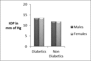
Figure 2: IOP in Diabetic group and Non Diabetic patients group Among the Diabetic patients the mean intraocular pressure among males (108 eyes) was 13.48 ±1.96 mm of Hg and females (32 eyes) was 13.69 ±1.69 mm of Hg. Among Non Diabetics the mean intraocular pressure in males (70 eyes) was 11.91 ±1.25 mm of Hg and females (30 eyes) was 11.93± 0.98 mm of Hg. There was no difference in the mean IOP between males and females in both groups. {Diabetic group (p= 0.6) and Non Diabetic group (p= 0.94)}Fig 3.
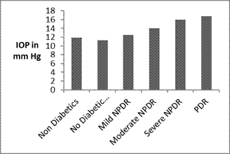
Figure 3: Association of Gender and IOP Among the Diabetic patients, mean IOP in patients with No Diabetic Retinopathy (8 eyes) was 11.25 ±1.04 mm of Hg, patients with Mild NPDR (62 eyes) was 12.52 ±1.25 mm of Hg, patients with Moderate NPDR (52 eyes) was 14.08 ± 1.43 mm of Hg, patients with Severe NPDR (08 eyes) was 16.00 ±0.00 mm of Hg, patients with PDR (10 eyes) was 16.8 ± 1.40 mm of Hg. There was significantly higher IOP in Mild, Moderate and Severe NPDR groups and PDR group than in Non Diabetic Group(p<0.001) One way ANOVA} Fig4.
Figure 4: IOP in different groups of Diabetic patients DISCUSSION There was no association between the Age and the IOP in our study which was similar to Beijing Eye Study.13 Whereas studies done by Hashemi H et al.18, David et al.19, Klein B E et al.7 and Bonomi L et al.20 found positive correlation between age and IOP. Contradicting to these studies was the Tajimi Study21 among the Japanese population which showed a negative correlation between age and IOP which was significant. In our study, there was no association between IOP and gender in both Diabetic and Non Diabetic group. This is similar to study done by Quigley HA 22 Our study reported that there was higher significant mean intraocular pressure in Diabetic patients than in Non Diabetic patients which was statistically significant. There are well documented studies 6-10 which shows positive relationship between IOP and Diabetes. There was significant increase in IOP as Diabetic Retinopathy progresses. Worldwide cause of irreversible blindness is Glaucoma and Diabetic Retinopathy.22 Elevated IOP is suggested as the major risk factor for Primary open angle Glaucoma 6-10 But few studies 11,14,22 have reported that the Higher IOP was associated with Diabetes but not with the development of Glaucoma. Various mechanisms are determined for the cause of higher intraocular pressure in patients with Diabetes. An epidemiological study done by Clark C V suggested the role of Genetic factor in ocular Hypertension.23 Study done by Mapstone et al.24 reported that the dysfunction of autonomic system may be the contributing factor for increase in the IOP. Mitchell Pet al.9 study suggested that osmotic gradient fluid shift into the intraocular space induced due to Elevated blood glucose level will elevate the IOP.
CONCLUSION Our study showed higher IOP in patients with Diabetes Mellitus than Non Diabetic patients. As IOP is a major risk factor for Glaucoma, it has to be monitored regularly for early detection in Diabetic patients. Early detection and treatment may reduce or help to limit ocular morbidity caused due to Glaucoma in Diabetic Patients.
REFERENCES
Policy for Articles with Open Access
|
|
||||||||||||||||||||
 Home
Home


