 Home
Home
|
Table of Content - Volume 16 Issue 2 - November 2020
Limbal conjuntival auto grafting for pterygium surgery with no suture no glue
R Niraimozhi1, S Nisar Ahamed2*
1Assistant Professor, 2Post Graduate, Department of Ophthalmology, K A P Vishwanatham Government Medical College, Mgmgh, Trichy,
Abstract Background: Limbal Conjunctival autografting in pterygium surgery without the use of sutures and fibrin glue is being practiced by some surgeons recently. The present study was undertaken to evaluate the outcome of the procedure Methods: This retrospective study was conducted by reviewing the records of thirty‑five consecutive cases of pterygium surgery without sutures and no glue, autolimbal conjunctival grafting done by a single surgeon from JAN1, 2019, to April 30,2019. two patient had nasal and temporal pterygium, and all other patients had primary nasal pterygium. Under peribulbar block, pterygium was excised and autolimbal conjunctival grafting was performed without sutures and no glue. Grafts were taken from the superotemporal bulbar conjunctiva of the same eye. Postoperative follow‑up was done on the 1st, 3rd and 7th postoperative day and then after 1 month and 2 months. Results: Patients who had graft edema, subconjunctival hemorrhage, dellen, graft recession or displacement during immediate postoperative period eventually settle down, and the cosmetic outcome was good in all patients. Except one in all other cases, grafts were Insitu. None of them had recurrence till the last follow‑up. Conclusion: Conjunctival limbal auto grafting with pterygium surgery without suture no glue takes minimal surgical time and no postoperative discomfort and suture related complication, economic and has better cosmetic outcome.
INTRODUCTION Pterygium is a Greek word meaning wing. It is a degenerative condition of the conjunctiva characterized by proliferation of subconjunctival tissue as a triangular fleshy mass or fold encroaching the cornea. Nasal pterygium is more common than temporal pterygium. The exact etiology is not known. exposure to ultraviolet rays is most common risk factors for the occurrence of pterygium. possibility of stem cell dysfunction induced by UV rays is related to formation of pterygium. other causes include continuous exposure to dry and dusty environment. Subconjunctival tissue undergoes elastotic degeneration of the collagen fibers of the substantia propria of conjunctiva with fibrovascular proliferation of granulation tissue under the epithelium which ultimately encroaches cornea and it causes redness, irritation, and visual disturbances due to irregular corneal astigmatism. If pterygium extends to the visual axis, it can block the vision altogether. Surgery is indicated when there is reduced vision due to astigmatism or encroachment of the visual axis, recurrent inflammation, and for cosmesis. Bare sclera excision of pterygium results in a significantly higher recurrence rate than excision accompanied by the use of certain adjuvants. conjunctival or limbal autograft is superior to amniotic membrane graft surgery in reducing the recurrence rate. Currently the best surgical option in terms of recurrence is conjunctival limbal autograft. In conjunctival autografting after pterygium excision, the graft is usually sutured or glued to the bed using fibrin glue to secure its position. suturing requires considerable skill from the surgeon associated with the prolonged operation time, postoperative discomfort, and suture related complication, with fibrin glue, there is a risk of hypersensitivity reaction and viral transmission and is expensive these problem lead to the development of sutureless and glue free conjunctival autografting technique. When tissue is wounded, blood comes in contact with collagen triggering blood platelets to begin secreting inflammatory in this technique. the conjunctival graft is placed onto the bed where the oozing blood clots and form a bioadhesive which secures the grafts in its position. the present study was undertaken to evaluate the outcome of the technique of sutureless and glue-free conjunctival autografting using patient’s own blood to secure the graft in place.
METHODS Medical records of 35 consecutive patients who underwent sutureless glue free conjunctival auto grafting with pterygium surgery done by a single sugeon at mahatma Gandhi govt. memorial hospital trichy between -JAN 01 TO APRIL 30 2019 were reviewed All patients had primary pterygium. out of 35 patient, 33 patients had nasal pterygium and 2 patient had primary double pterygium both (nasal and temporal) Criteria for patient selection were as follows: Inclusion criteria: People of age group >20 years, True pterygium, Patients with other comorbid conditions like diabetes and hypertension Exclusion criteria: Pediatric patients. Patients with fundus pathology. Glaucoma patients. Pseudopterygium. Injuries of eye. Suspected ocular surface squamous neoplasia. The patients who had asymmetrical pterygium in both eyes. SURGICAL PROCEDURE Under peribulbar block after placing the speculum, the eye was painted with povidone iodine 10% solution then drapped, pterygium area was mopped dry. pterygium was excised starting from the body and the head was stripped off by reverse avulsion the remnants were cleared using took’s knife. after mopping dry, graft area was marked 1mm larger than bare scleral area in both dimensions. Conjunctiva was ballooned by injecting saline subconjunctival. Conjunctival graft was dissected carefully avoiding tenon’s capsule and without button holes using non toothed forceps and round tipped scissors. after clearing the recipient bed of excess blood, the graft was placed on the bare scleral area in such a way that limbal side of the graft overlies the limbus of the scleral bare area. once properly positioned, the graft was left undisturbed for 5mins. After mopping excess blood antibiotic eye ointment was applied. speculum was removed gently without disturbing the graft and eye patch applies. patch was removed after 24hr, and all patient were examined under slit lamp after 24hr and followed up after 1, 3 and 7 day and 1 month for graft edema, subconjunctival hemorrhage, graft retraction, dellen, and recurrence of pterygium.
RESULTS A total of 35 Eyes of 35 patients with primary pterygium were included in this study. The mean age of the studied population was 40 ± 1. The male and female distribution was 6:29[Table1]. Out of 35 eyes, 5 had subconjunctival hemorrhage, 2 had graft edema, 2 had dellen , 1 had graft loss, and 2 had graft recession [Table3]. At 2 months follow‑up, no recurrence was found. All patient had good cosmetic outcome. Table 1: Gender distribution
Table 2: types of pterygium
Table3: outcome of surgery
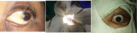
Figure 1: Pre operative image nasal pterygium left eye Figure 2: Intraoperative Image Figure 3: Post-operative image DISCUSSION Pterygium surgery should ideally be easy to perform, cost effective, and cosmetically acceptable with no recurrence. Several surgical techniques have evolved over the years. Bare scleral technique was the one that was introduced first. This has been abandoned due to high recurrence rate.9 Application of intraoperative mitomycin C had serious complications with scleral necrosis/ melting and ocular perforation.7 Currently conjunctival autografting is the standard procedure for pterygium surgery with low recurrence rate. 12 The graft was usually fixed to the scleral bed using sutures. However, this procedure involves prolonged surgical time, postoperative discomfort, and other suture‑related complications. To overcome these difficulties, fibrin glue application to fix the graft in place was developed. 6 However, this has disadvantages such as higher cost, anaphylactic reaction, and risk of viral transmission. To overcome these drawbacks, conjunctival autografting using patient’s own blood to secure the graft in place has been introduced recently. This technique has overcome several disadvantages encountered with earlier methods. The technique is easier to perform with less discomfort to patients and is cost‑effective. In our series, one case had lost graft due to excessive bleeding which could be attributed to surgeon’s learning curve. Patients who had dellen and graft recession eventually settled down. The cosmetic outcome was good in all the patients. No recurrence was found, but the follow‑up period was only 4 months. The result was comparable to other studies using similar technique. 10 Sutureless glue‑free conjunctival autografting with pterygium surgery is an excellent technique easy to perform, with short surgical time, good cosmesis, and no recurrence. Earlier studies have shown that calcium accelerates the rate of fibrin monomer polymerization, thereby decreasing the time required to form clot. 13 Studies may be undertaken in the future to find whether addition of calcium to the scleral bed helps in faster and better adherence of the conjunctival graft. Conclusion Sutureless glue‑free conjunctival autografting in pterygium surgery takes short surgical time, is economic, and has good cosmetic outcome.
REFERENCES
|
|
 This work is licensed under a Creative Commons Attribution-NonCommercial 4.0 International License.