 Home
Home
|
Table of Content - Volume 17 Issue 3 - March 2021
A study of treatment modalities in early post-operative endophthalmitis
Santosh Y Tupdikar
Assistant Professor, Department of Ophthalmology, MIMSR Medical College, Latur, Maharashtra, INDIA. Email: skyt55@gmail.com
Abstract Background: endophthalmitis though rare, still remains the most dramatic and devastating complications of cataract and other intra-ocular surgeries. Aim and Objective: To study treatment modalities in early post-operative endophthalmitis. Methodology: This was a cross –sectional study in the patients with total 60 Eyes were studied over period of 2 Years i.e. 2004 to 2006, All patients with signs and symptoms suggestive of endopthalmitis following intra-ocular surgery were enrolled into the study. The data was collected by pre-tested, semi-structured questionnaire, the data was analyzed by Chi-square test and calculated by SPSS 19 version. Result: Majority of the patients undergone Pars plana Vitrectomy i.e. 46.66%, Aggressive Topical and Intra-vitreal injection undergone 43.33%, Aggressive Topical Therapy Only in8.33%. As per the Associated other interventions majority of the patients undergone Anterior chamber wash, In 62.26%, Penetrating Keratoplasty in 9.43%, Implant removal in 5.66%, Enucleation in 3.77%, IOFB Removal in 1.88%. Majority of patients with Initial Visual Acuity were having Light perception in- 26; Vision (1/60)-21, Counting Fingers -5 and Final Acuity after all the treatment majority of the patients were having Vision (1/60)- were 12, Vision (>6/60) in 10 this observed difference was statistically significant (χ2 =10.17, df=4, p<0.38). Conclusion: It can be concluded from our study that though the Endopthamitis is dreaded complication but if it is detected early and treated with appropriately is having good outcome. Key words: endophthalmitis, IOFB (Foreign Intra Ocular Body), IOL (Intra Ocular Lens).
INTRODUCTION endophthalmitis though rare, still remains the most dramatic and devastating complications of cataract and other intra-ocular surgeries. It is the severest and vision threating form of ocular infection, the mere mention of which invokes the fear of God in the mind of any practicing Opthalmologist. Endophthalmitis may also follow penetrating variety ocular trauma, microbial keratitis and endogeneous infection. The phenomenal variety of its presentation ranging from smoldering, indulent process to a Virulence suppurative one can be source of Diagnostic confusion. The management of patents with endophthalmitis is even more probelemetic to the Ophthalmologists who sees it in frequently the conflicting recommendations with regard to culture techniques, antimicrobial Treatment and surgery are indeed unsettling. So in our study we have studied the different treatment modalities in early post-operative endophthalmitis and it’s final outcome.
METHODOLOGY This was a cross–sectional study in the patients with total 60 Eyes were studied over period of 2 Years i.e. 2004 to 2006, All patients with signs and symptoms suggestive of endopthalmitis following intra-ocular surgery were enrolled into the study. Most of the patients had undergone cataract surgery or a secondary IOL implantation, through a few were cases of endopthalmitis following other surgeries like trabeculectomy, Keratoplasty and Vitrectomy. The commonest presenting complaint was decreased visual acuity but patients also presented with other complain such conjunctival injection, ciliary congestion, Ocular pain and headache, watering and purulent discharge. All the patients entering the study were subjected to through examination. The data was collected by pre-tested, semi-structured questionnaire, the data was analyzed by Chi-square test and calculated by SPSS 19 version.
RESULT Table 1: Distribution of the patients as per the Age
The majority of the patients were in the age group of 41-50, 51-60 were 14, followed by 61-70 were 13, 31-40 were 8, >70 were 7, 21-30-3, 11-20 were 1.
Table 2: distribution of the patients as per the sex
The majority patients were Male i.e. 33 and Female were 27.
Table 3: Distribution of the patients as per the Primary Interventions
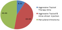
Majority of the patients undergone Pars plana Vitrectomy i.e. 46.66%, Aggressive Topical and Intra-vitreal injection undergone 43.33%, Aggressive Topical Therapy Only in8.33%.
Graph 1: Distribution of the patients as per the Primary Interventions Table 4: Distribution of the patients as per the Associated other interventions
As per the Associated other interventions majority of the patients undergone Anterior chamber wash In 62.26%, Penetrating Keratoplasty in 9.43%, Implant removal in 5.66%, Enucleation in 3.77%, IOFB Removal in 1.88%.
Table 5: Distribution patients as per the Initial Versus Final Visual Acuity
(χ2 =10.17, df=4, p<0.38*) Majority of patients with Initial Visual Acuity were having Light perception in- 26; Vision (1/60)-21, Counting Fingers -5 and Final Acuity after all the treatment majority of the patients were having Vision (1/60)- were 12, Vision (>6/60) in 10 this observed difference was statistically significant (χ2 =10.17, df=4, p<0.38).
DISCUSSION Acute postoperative endophthalmitis following cataract surgery is a dreaded complication. Fortunately, the incidence has declined in recent times after changes in surgical techniques, sterilization procedures, and better understanding of the risk factors.1,2,3,4,5,6,7 The global reported incidence of postcataract endophthalmitis ranges from 0.02% to 0.26%.1,2,3,4,5,6,8,9,10,11 There is good amount of data on the incidence, risk factors, and outcomes of postcataract endophthalmitis in the Western hemisphere.12 Over the past decade, there has been a decline in the incidence of postoperative endophthalmitis, owing to the improvement of modern surgery, instrumentation, sterility, and prophylactic antibiotics. Generally accepted as approximately 10% at the beginning of the century,1 the incidence today ranges from 0.3% (prospective study realised in 1989 in France)2 down to 0.07% (retrospective American study reporting data from 1984 to 1989).3 Incidence seems to depend on the type of surgery. After extracapsular lens extraction (ECLE) or phacoemulsification and intraocular lens (IOL),34 it is between 0.07% and 0.12% After secondary IOL implantation, it is higher (0.3%), probably related to greater manipulation. Endophthalmitis immediately following trabeculectomy is rare (0.6%), with an apparent higher incidence of late onset endophthalmitis (1.8%) occurring from 3 months to 27 years postoperatively.5 Of particular concern is the high incidence of rapidly progressing and devastating late onset endophthalmitis after the use of the antimetabolites 5-fluorouracil (5–8%)8 and mitomycin C (MMC) (2.7–3%).9 In our study we have found The majority of the patients were in the age group of 41-50, 51-60 were 14, followed by 61-70 were 13, 31-40 were 8 , >70 were 7 , 21-30-3, 11-20 were 1 The majority patients were Male i.e. 33 and Female were 27 . Majority of the patients undergone Pars plana Vitrectomy i.e. 46.66%, Aggressive Topical and Intra-vitreal injection undergone 43.33%, Aggressive Topical Therapy Only in8.33%. As per the Associated other interventions majority of the patients undergone Anterior chamber wash In 62.26%, Penetrating Keratoplasty in 9.43%, Implant removal in 5.66%, Enucleation in 3.77%, IOFB Removal in 1.88%. Majority of patients with Initial Visual Acuity were having Light perception in- 26; Vision (1/60)-21, Counting Fingers -5 and Final Acuity after all the treatment majority of the patients were having Vision (1/60)- were 12, Vision (>6/60) in 10 this observed difference was statistically significant (χ2 =10.17, df=4, p<0.38). This is similar with the study done by Bajimaya et al. (2010)20 and Carrim et al. (2009)21. About 74% (14/19) of their patients had achieved best corrected visual acuity better than or equal to 6/60. And with Gautam P et al. 22 they found The best corrected visual acuity of 6/9 was achieved in 2 patients, 5 had 6/18, 2 had 6/60 and 2 had 5/60 at the end of eight weeks.
CONCLUSION It can be concluded from our study that though the Endopthamitis is dreaded complication but if it is detected early and treated with appropriately is having good outcome.
REFERENCES
Policy for Articles with Open Access: Authors who publish with MedPulse International Journal of Pediatrics (Print ISSN: 2579-0897) (Online ISSN: 2636-4662) agree to the following terms: Authors retain copyright and grant the journal right of first publication with the work simultaneously licensed under a Creative Commons Attribution License that allows others to share the work with an acknowledgement of the work's authorship and initial publication in this journal. Authors are permitted and encouraged to post links to their work online (e.g., in institutional repositories or on their website) prior to and during the submission process, as it can lead to productive exchanges, as well as earlier and greater citation of published work.
|
|
 This work is licensed under a Creative Commons Attribution-NonCommercial 4.0 International License.