 Home
HomeOfficial Journals By StatPerson Publication
|
Table of Content - Volume 10 Issue 1 - April 2019
A study of role of obesity in the chondral lesions of isolated medial meniscus tears at tertiary health care centre
Vaibhav Bhadbhade1, Channabasava2*, Dayanand M3, Ramaligaiah A4
1,2Senior Resident, 3Assistant Professor, 4Professor, Department of Orthopaedics, BMCRI, Bangalore, Karnataka, INDIA. Email: vaibhavrb9@gmail.com
Abstract Background: A medial meniscus tear is more common than a lateral meniscus tear, because it is firmly attached to the deep medial collateral ligament and the joint capsule. The medial meniscus absorbs up to 50% of the shock of the medial compartment so it is susceptible to injury. Meniscal injury is a major cause of functional impairment of the knee. Aim and objective: To study the role of obesity with isolated chondral lesions of medial meniscus Methodology: Present study was prospective study carried out in patients with isolated medial meniscus tear. Data was collected with pretested questionnaire. Data included sociodemographic data, BMI, detailed history and Clinical examination. Diagnostic arthroscopy was performed to evaluate abnormal findings. The severity of chondral damage was determined by the ICRS grade and the number of compartments involved. Data was analysed with appropriate statistical tests. Results and discussion: Majority of the patients were from age group of > 50 years (55%) followed by 41-50 years (22%). Incidence increases as age increases in females. We observed that chondral score was significantly affected by BMI (P value <0.05) Key Word: obesity, chondral lesions.
INTRODUCTION Osteoarthritis (degenerative arthritis, osteoarthrosis, artrosis, OA) is a slowly progressive joint disease, which may lead to local destruction of the joint. Osteoarthritis was listed among the top 10 of the global disease burden according to the World Health Organization1 .It is known that the menisci are important in the function of the knee joint. Tibiofemoral load transmission, shock absorption, and lubrication are the main functions of mensci.2-4 The menisci compensate for significant incongruity between the femoral and tibial articulating surfaces. The human menisci transmit 30–55% of the load in a standing position.2 Injuries to the menisci are one of the most common knee injuries. They can occur in isolation or with associated injuries to knee ligaments. The meniscal tear is commonly due to internal rotation of the femur as the flexed knee moves toward an extended position. The meniscus usually splits in a longitudinal direction. The central part of the meniscus may dislocate centrally and cause locking of the knee. A thorough physical exam, patient history, and the use of magnetic resonance imaging (MRI) are the primary means of diagnosing meniscal injuries. Arthroscopic treatment involving removing or repairing the meniscal tear is the primary method of treatment. Prevalence of obesity is increasing worldwide. Developed as well as developing countries showed increased prevalence over last decade. Obesity contributes to various non communicable diseases. Obesity has effects on osteoarthiritis also.According to study by Reyes C et al5 when Compared with patients with normal weight, a body mass index (BMI) between 25 and 30 increases the risk of osteoarthritis by a factor of 2, a BMI between 30 and 35 by a factor of 3, and a BMI above 35 by a factor of 5. Present study was done to find out the role of obesity isolated chondral lesions of medial meniscus.
METHODOLOGY Present study was a prospective study that was conducted in 100 patients over a period of one year from July 2017 to June 2018 in a tertiary care centre banaglore medical college and research institute, bangalore. Study population were patients with isolated medial meniscus tear. Inclusion criteria
Exclusion criteria
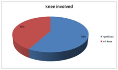
Study was approved by ethical committee. A valid written consent was taken from patients after explaining study to them. Data was collected with pretested questionnaire. Data included socio-demographic data like age, sex, Body Mass Index of the patients and detailed history. Clinical examination done. All patients undergone pre operative assessment before surgery. Patients underwent arthoscopy. Surgery was done under general anesthesia. Standard anterolateral and anteromedial knee portals were used. Diagnostic arthroscopy was performed to evaluate abnormal findings. Medial meniscal tears were trimmed to a stable rim. At surgery, cartilage lesions were probed, measured and then graded according to the international cartilage repair society (ICRS) classification.6 The severity of chondral damage was determined by the ICRS grade and the number of compartments involved. If two or more compartments had an ICRS score of at least 2 then the total score was raised by 1 point (i.e. the chondral score range was 0-5). Body mass index was considered normal at 18.5-24.9 kg/m2, overweight at 25-29.9 kg/m2 and obese if ≥30 kg/m2 values, according to WHO guidelines. RESULTS Total 100 patients were studied. Table one shows distribution of patients according to age group and sex.In our study we found that majority of the patients were from age group of > 50 years (55%) followed by 41-50 years (22%). 18-30 years age group has only 8 patients.In our study we found that majority of the patients were males (68%). Overall male: female ratio was 2.1:1. Incidence of meniscal tear was more in males upto the age of 50 years. Incidence was almost same after the age of 50 years in both male and female.In age group of 18-30 years no female was suffered from meniscal tear. Incidence increases as age increases in females. In our study 58% patients had right side medial meniscus tear and 42% had left sided tear. Table 3 shows distribution of patients according to age group mean BMI and Mean Chondral score.Mean age of patients was 50.23±3.41 years (18-82 years) and an mean BMI of 27.35±2.3 (19.2-37.2) kg/m2.Females had higher BMI [mean 28.7 kg/m2 (range 19.4-37.2 kg/m2)] than males (mean 26.4 kg/m2 (range 19.8-36.9 kg/m2). There were 40 patients with no chondral lesions, 6 patients with a chondral score of 1, 19 patients with chondral score of 2, 15 patients with chondral score of 3, 17 patients with chondral score of 4 and only 3 patients with chondral score of 5 were seen.By applying the ANNOVAwe confirmed that chondral score was significantly affected byBMI (P value <0.05)
Table 1: Distribution of patients according to age group and sex
Figure 1: distribution of patients according to side of knee involved
Table 2: Distribution of patients according to age group mean BMI and Mean Chondral score
DISCUSSION In our study we found that majority of the patients were from age group of > 50 years (55%) followed by 41-50 years (22%). In younger age, the biochemical composition of the joint cartilage can absorb the mechanical stresses around the knee.The degenerative process accelerates from the 5th decade of life. Similarily Camanho et al 7found arthroscopic knee changes were more common inaverage age of50-59 years (34.7%) among 435 patients .Overall male: female ratio was 2.1:1. Incidence of meniscal tear was more in males upto the age of 50 years. Incidence increases as age increases in females. Previous studies showed demographic sex differences in meniscal tears. Demographic sex differences were also found in previous works on meniscal tears.8-10 In our study we found that chondral score was significantly affected byBMI (P value <0.05)A higher body mass cause accelerated damage to the joints by higher tibiofemoral compressive and shear forces11. Obese individuals spend a greater portion of the gait cycle in the stance phase and have greater adduction moment curves increasing the cumulative load that further contributes to joint damage.12 Significant associations were demonstrated between increased BMI and meniscal lesions in both genders, including obese and overweight adults 13.
CONCLUSION BMI (obesity) affects degree of chondral lesions in isolated medial meniscus tear.
REFERENCES
|
|
 This work is licensed under a Creative Commons Attribution-NonCommercial 4.0 International License.