 Home
Home|
Table of Content - Volume 15 Issue 3 - September 2020
Functional outcome of surgical management of distal radius fracture by LCP
Anwar Hamzath AK1, Reneesh U P2*
1,2Assistant Professor, Assistant Professor, Department of Orthopedics, DMWIMS, Naseera Nagar, Meppadi, Wayanad, Kerala, INDIA. Email: drreneesh@gmail.com
Abstract Background: Distal Radius fractures are one of the commonest injuries that affect the elderly population and also the young. Distal Radius fracture is a leading cause of hospital admissions in elderly people. The number of such admissions is on a raise because of increasing life span and sedentary habits. Conservative methods of treatment results in malunion with shortening and limitation of wrist movement. This study is done to analyze the surgical management of Distal Radius fractures using Locking Compression Plate. Methods: This is a prospective study of 30 cases of fresh Distal Radius fractures admitted tohospital.. Cases were taken according to inclusion and exclusion criteria i.e., patients with Distal Radius fracture above the age of 18yrs. Medically unsuitable and patients not willing for surgery were excluded from the study. Results: In our series of 30 cases there were 19(63.33%) male and 11(36.66%) female, RTA was the most common mode of injury accounting for 21 (70%) cases and fall accounted for 7 (23.33%)cases. In our study 27 (90%) cases were closed fractures and 3 (10%) cases were open fractures of Gustilo type I. In the present series, commonest duration between trauma and surgery was 2 to 4 days. 24 (80%) underwent surgery within 2 to 4 days after trauma. The average duration of hospital stay in the present study is 11.73 days. Conclusion: From this sample study, we consider that LCP is an excellent implant for the treatment of Distal Radius fractures. The terms of successful outcome include a good understanding of fracture biomechanics, proper patient selection, good preoperative planning, accurate instrumentation, good image intensifier. Key Words: LCP; Volar Plating; Distal Radius.
INTRODUCTION The past decade has witnessed widespread enthusiasm for operative treatment of distal radial fractures. A number of factors have contributed to this changing perspective. Among these include a greater recognition of the wide variation in fracture patterns. Recognition of this unique anatomical orientation and patterns of articular injury has led to the development of a method of operative treatment coined fragment-specific fixation, through which small strategically-placed implants are used to support the critical structural components of the fracture1. Main aim is to achieve anatomic union, early range of motion2. Another factor that has led to the operative treatment of certain fractures involves those injuries associated with intercarpal injury. Fracture patterns involving compression or shearing of the radial styloid and/or scaphoid facet of the distal radius are recognized to have the potential of accompanying injuries to the scapholunate ligament as well as the lunate facet and the lunotriquetral ligament. Associated instability of the distal radioulnar joint (DRUJ) has also stimulated more operative intervention. Volar plating provides easier surgical access and protects tendon sheath by the pronator quadratus3. It provides a stable construct and early mobilization4. DRUJ instability, especially in the presence of a large ulnar-styloid fracture, is best treated by operative fixation of the ulnar styloid. MATERIALS AND METHODS This is a prospective clinical study of surgical treatment of distal end of radius fracture by LCP. In all, a total of 30 cases were evaluated and assessed. All the patients underwent treatment, as per a specific treatment plan. All the patients were initially assessed in the out-patient department or casualty according to their presentation and then they underwent a detailed evaluation of their hemodynamics(table 1) , neurological status and other injuries if associated with trauma5(table 2) The patients were interviewed; their epidemiological, historical, subjective and physical findings were noted(table 3). After initial investigations and hemodynamic stabilization, patients were Exclusion criteria were –
SURGICAL TECHNIQUE- Under general or regional anesthesia a volar approach with an 8-10cm skin incision along the radial side of flexor carpi radialis(FCR) was used. After splitting the forearm fascia, the FCR tendon, flexor tendons and median nerve was retracted to the ulnar side and the pronator quadratus muscle was detached from the radius8. Reduction was performed under direct vision and confirmed with an image intensifier and the bone was fixed temporarily with Kirschner wires. Then the plate was inserted and fixed in the gliding hole9. Correct positioning was verified using an image intensifier. After placement of the plate the detached pronator quadratus muscle was reattached with absorbable sutures. After the wound closure a compressive dressing and a splint were applied. Postoperatively the wrist was immobilized for 4 weeks with application of a short arm slab for 1 week and short arm cast for 3 weeks. Range of motion exercises for the wrist were started at 4 weeks after surgery. EVALUATION OF OUTCOME The patients were followed up for minimum of 54 wks. Clinical, radiological and functional reviews were performed at periodic intervals. OBSERVATIONS AND RESULTS The study comprised a total of thirty patients of fractures of the distal radius. Table 1: Type of Fracture
Table 2: Time interval between trauma and surgery
Table 3: Stay in Hospital
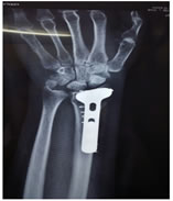
Figure 1: 56 year old female Figure 2: 48 year old male Figure 3: 59 year old male Figure 4: 61 year old female Figure 5: Age Incidence Figure 6: Sex Incidence Figure 7: Side Affected
ANATOMICAL SCORE OF HEALED FRACTURE The scoring was done according to the Sarmiento’s modification of Lind Storm Criteria. Anatomically 26 patients (86.67%) had excellent restoration of anatomy, 3 patients (10%) had good restoration and 1 had fair (3%) restoration of anatomy. Thus 96% patients had excellent to good alignment of fragments and good reduction could not be achieved in 3% patients resulting in fair or poor results. FUNCTIONAL END RESULT OF HEALED FRACTURE The scoring of healed fracture was done as according to the Demerit point system of Gartland and Werley with Sarmiento et al.’s modification. Functionally 24 patients (80%) had excellent, 5 good (17%) and 1 patients had fair (3%) restoration of functions. Poor function correlated with residual displacement and poor patient compliance.
DISCUSSION The treatment has changed over the previous two decades10. Few prospective studies confirm the benefit of this increasingly aggressive operative approach and surgical management of fracture of the distal radius11. Studies showed plates allow early mobilization and good support. Majority of the studies have used subjective tools for measuring quality of life. Implants allowed limited fixation of fracture components and minimization of prior problems associated with larger nonspecific implants12. Volar implants with locked head screws provide angular stability even in osteoporotic patients. Stability is enhanced by placing the distal screws in a subchondral position especially when stabilizing a dorsally displaced fracture from a volar approach13. A number of options regarding internal fixation are dependent upon the fracture pattern and available implants. The indications for bone graft, bone substitute will be based upon whether a defect exists under an articular fragment and the stability of the internal fixation14. The development of implants with locked screw heads, which creates angular stable fixation, has substantially decreased the requirement for bone graft. Following internal fixation of the radius fracture, the stability of the distal radioulnar joint should be assessed in every patient. This is best performed by attempting to displace the distal ulna from the sigmoid notch of the radius in forearm pronation, neutral and supination. If the ulna head can be displaced completely, the possibility of instability should be considered. Volar plating provide stable fixation and avoids many complication15. The treatment goal of these fractures is a wrist that provides sufficient pain-free motion and stability without the propensity for future degenerative changes. Although this goal may be easily accepted, there is very little consistency regarding the radiographic features that will afford this result. It seems rational to use LCP for fracture radius as an effective treatment method in terms of early functional mobilization16.
CONCLUSION Functional and Anatomical results, suggests that stabilizing the fracture fragments with volar plate and screws in the management of the fractures of distal radius, is an effective method to maintain the reduction till union and prevent collapse of the fracture fragments, even when the fracture is grossly comminuted/intra-articular/unstable and/or the bone is osteoporosed. The technique emphasizes that open reduction and internal fixation with volar plating has excellent functional outcome with minimal complications thus proving that it is the prime modality of treatment for distal radius fractures. The procedure is applicable for AO types A, B and C fractures of the distal radius, in young patients with a good bone stock as well as in elderly osteoporotic patients.
REFERENCES
.
Policy for Articles with Open Access: Authors who publish with MedPulse International Journal of Forensic Medicine (Print ISSN: 2579-0935) (Online ISSN: 2636-4735) agree to the following terms: Authors retain copyright and grant the journal right of first publication with the work simultaneously licensed under a Creative Commons Attribution License that allows others to share the work with an acknowledgement of the work's authorship and initial publication in this journal. Authors are permitted and encouraged to post links to their work online (e.g., in institutional repositories or on their website) prior to and during the submission process, as it can lead to productive exchanges, as well as earlier and greater citation of published work.
|
|


 This work is licensed under a Creative Commons Attribution-NonCommercial 4.0 International License.